 Movie
Movie Controller

[English] 日本語
 Yorodumi
- PDB-3qip: Structure of HIV-1 reverse transcriptase in complex with an RNase... -
+ Open data

- Basic information
| Entry | Database: PDB / ID: 3qip | ||||||
|---|---|---|---|---|---|---|---|
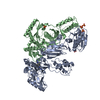
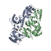
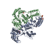
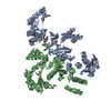
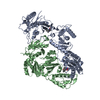
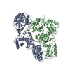
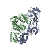
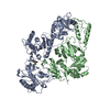
| Title | Structure of HIV-1 reverse transcriptase in complex with an RNase H inhibitor and nevirapine | ||||||
 Components Components |
| ||||||
 Keywords Keywords | TRANSFERASE/TRANSFERASE INHIBITOR /  HIV / reverse transcriptase / RNase H / polymerase / nuclease / TRANSFERASE-TRANSFERASE INHIBITOR complex HIV / reverse transcriptase / RNase H / polymerase / nuclease / TRANSFERASE-TRANSFERASE INHIBITOR complex | ||||||
| Function / homology |  Function and homology informationintegrase activity / Integration of viral DNA into host genomic DNA / Autointegration results in viral DNA circles / Minus-strand DNA synthesis / Plus-strand DNA synthesis / Uncoating of the HIV Virion / 2-LTR circle formation / Vpr-mediated nuclear import of PICs / Early Phase of HIV Life Cycle / Integration of provirus ...integrase activity / Integration of viral DNA into host genomic DNA / Autointegration results in viral DNA circles / Minus-strand DNA synthesis / Plus-strand DNA synthesis / Uncoating of the HIV Virion / 2-LTR circle formation / Vpr-mediated nuclear import of PICs / Early Phase of HIV Life Cycle / Integration of provirus / APOBEC3G mediated resistance to HIV-1 infection / Binding and entry of HIV virion / viral life cycle / Assembly Of The HIV Virion / HIV-1 retropepsin / : / Budding and maturation of HIV virion / retroviral ribonuclease H / exoribonuclease H / : / exoribonuclease H activity / protein processing / host multivesicular body / RNA-directed DNA polymerase / viral genome integration into host DNA / viral penetration into host nucleus / establishment of integrated proviral latency / RNA-directed DNA polymerase activity / Transferases; Transferring phosphorus-containing groups; Nucleotidyltransferases / RNA-DNA hybrid ribonuclease activity / peptidase activity / viral nucleocapsid / DNA recombination / Hydrolases; Acting on ester bonds / DNA-directed DNA polymerase / aspartic-type endopeptidase activity / DNA-directed DNA polymerase activity / symbiont entry into host cell / symbiont-mediated suppression of host gene expression / lipid binding / host cell nucleus / host cell plasma membrane / virion membrane / structural molecule activity / DNA binding / RNA binding / zinc ion binding / membrane / identical protein binding Function and homology informationintegrase activity / Integration of viral DNA into host genomic DNA / Autointegration results in viral DNA circles / Minus-strand DNA synthesis / Plus-strand DNA synthesis / Uncoating of the HIV Virion / 2-LTR circle formation / Vpr-mediated nuclear import of PICs / Early Phase of HIV Life Cycle / Integration of provirus ...integrase activity / Integration of viral DNA into host genomic DNA / Autointegration results in viral DNA circles / Minus-strand DNA synthesis / Plus-strand DNA synthesis / Uncoating of the HIV Virion / 2-LTR circle formation / Vpr-mediated nuclear import of PICs / Early Phase of HIV Life Cycle / Integration of provirus / APOBEC3G mediated resistance to HIV-1 infection / Binding and entry of HIV virion / viral life cycle / Assembly Of The HIV Virion / HIV-1 retropepsin / : / Budding and maturation of HIV virion / retroviral ribonuclease H / exoribonuclease H / : / exoribonuclease H activity / protein processing / host multivesicular body / RNA-directed DNA polymerase / viral genome integration into host DNA / viral penetration into host nucleus / establishment of integrated proviral latency / RNA-directed DNA polymerase activity / Transferases; Transferring phosphorus-containing groups; Nucleotidyltransferases / RNA-DNA hybrid ribonuclease activity / peptidase activity / viral nucleocapsid / DNA recombination / Hydrolases; Acting on ester bonds / DNA-directed DNA polymerase / aspartic-type endopeptidase activity / DNA-directed DNA polymerase activity / symbiont entry into host cell / symbiont-mediated suppression of host gene expression / lipid binding / host cell nucleus / host cell plasma membrane / virion membrane / structural molecule activity / DNA binding / RNA binding / zinc ion binding / membrane / identical protein bindingSimilarity search - Function | ||||||
| Biological species |  HIV-1 M:B_HXB2R (virus) HIV-1 M:B_HXB2R (virus) | ||||||
| Method | X-RAY DIFFRACTION / SYNCHROTRON / MOLECULAR REPLACEMENT / Resolution: 2.0926 Å | ||||||
 Authors Authors | Lansdon, E.B. / Kirschberg, T.A. | ||||||
 Citation Citation | Journal: Antimicrob.Agents Chemother. / Year: 2011 Title: Structural and Binding Analysis of Pyrimidinol Carboxylic Acid and N-Hydroxy Quinazolinedione HIV-1 RNase H Inhibitors. Authors: Lansdon, E.B. / Liu, Q. / Leavitt, S.A. / Balakrishnan, M. / Perry, J.K. / Lancaster-Moyer, C. / Kutty, N. / Liu, X. / Squires, N.H. / Watkins, W.J. / Kirschberg, T.A. | ||||||
| History |
|
- Structure visualization
| Structure viewer | Molecule: MolmilJmol/JSmol |
|---|
- Downloads & links
-Download
| PDBx/mmCIF format | 3qip.cif.gz | 218.4 KB | Display | PDBx/mmCIF format |
|---|---|---|---|---|
| PDB format | pdb3qip.ent.gz | 172.2 KB | Display | PDB format |
| PDBx/mmJSON format | 3qip.json.gz | Tree view | PDBx/mmJSON format | |
| Others |  Other downloads Other downloads |
-Validation report
| Arichive directory | https://data.pdbj.org/pub/pdb/validation_reports/qi/3qipftp://data.pdbj.org/pub/pdb/validation_reports/qi/3qip | HTTPS FTP |
|---|
-Related structure data
| Related structure data | 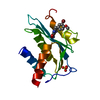 3qinC 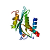 3qioC 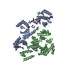 1vrtS S: Starting model for refinement C: citing same article ( |
|---|---|
| Similar structure data |

-Links
 PDBj
PDBj


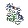
- Assembly
| Deposited unit | 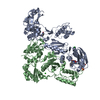
| ||||||||
|---|---|---|---|---|---|---|---|---|---|
| 1 |
| ||||||||
| 2 | 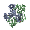
| ||||||||
| Unit cell |
| ||||||||
| Components on special symmetry positions |
|
-Components
-Protein , 2 types, 2 molecules AB
| #1: Protein | Mass: 64562.949 Da / Num. of mol.: 1 / Fragment: p66 subunit (UNP residues 588-1147) Source method: isolated from a genetically manipulated source Source: (gene. exp.) HIV-1 M:B_HXB2R (virus) / Gene: gag-pol / Plasmid: pET14b / Production host:  Escherichia coli (E. coli) / Strain (production host): BL21(DE3) Escherichia coli (E. coli) / Strain (production host): BL21(DE3)References: UniProt: P04585, HIV-1 retropepsin, RNA-directed DNA polymerase, DNA-directed DNA polymerase, retroviral ribonuclease H |
|---|---|
| #2: Protein | Mass: 51399.047 Da / Num. of mol.: 1 / Fragment: p51 subunit (UNP Residues 588-1027) Source method: isolated from a genetically manipulated source Source: (gene. exp.) HIV-1 M:B_HXB2R (virus) / Gene: gag-pol / Plasmid: pET 14b / Production host: Escherichia coli (E. coli) / Strain (production host): BL21(DE3)References: UniProt: P04585, HIV-1 retropepsin, RNA-directed DNA polymerase, DNA-directed DNA polymerase, retroviral ribonuclease H |
-Non-polymers , 6 types, 426 molecules 


| #3: Chemical | ChemComp-NVP / Nevirapine Mass: 266.298 Da / Num. of mol.: 1 / Source method: obtained synthetically / Formula: C15H14N4O / Comment: medication, antiretroviral*YM Mass: 266.298 Da / Num. of mol.: 1 / Source method: obtained synthetically / Formula: C15H14N4O / Comment: medication, antiretroviral*YM | ||||||||
|---|---|---|---|---|---|---|---|---|---|
| #4: Chemical |  Mass: 54.938 Da / Num. of mol.: 2 / Source method: obtained synthetically / Formula: Mn Mass: 54.938 Da / Num. of mol.: 2 / Source method: obtained synthetically / Formula: Mn#5: Chemical | Sulfate Mass: 96.063 Da / Num. of mol.: 2 / Source method: obtained synthetically / Formula: SO4 Mass: 96.063 Da / Num. of mol.: 2 / Source method: obtained synthetically / Formula: SO4#6: Chemical | Chloride Mass: 35.453 Da / Num. of mol.: 2 / Source method: obtained synthetically / Formula: Cl Mass: 35.453 Da / Num. of mol.: 2 / Source method: obtained synthetically / Formula: Cl#7: Chemical | ChemComp-P4Y / |  Mass: 361.351 Da / Num. of mol.: 1 / Source method: obtained synthetically / Formula: C20H15N3O4 Mass: 361.351 Da / Num. of mol.: 1 / Source method: obtained synthetically / Formula: C20H15N3O4#8: Water | ChemComp-HOH / | WaterMass: 18.015 Da / Num. of mol.: 418 / Source method: isolated from a natural source / Formula: H2O |
-Experimental details
-Experiment
| Experiment | Method: X-RAY DIFFRACTION / Number of used crystals: 1 |
|---|
- Sample preparation
| Crystal | Density Matthews: 3.04 Å3/Da / Density % sol: 59.52 % |
|---|---|
| Crystal grow | Temperature: 293 K / Method: vapor diffusion, hanging drop / pH: 6.5 Details: 1.3M (NH4)2SO4, 5mM sodium malonate, and 100mM cacodylate pH 6.5, VAPOR DIFFUSION, HANGING DROP, temperature 293K |
-Data collection
| Diffraction | Mean temperature: 100 K |
|---|---|
| Diffraction source | Source: SYNCHROTRON / Site: ALS  / Beamline: 5.0.2 / Wavelength: 1 Å / Beamline: 5.0.2 / Wavelength: 1 Å |
| Detector | Type: ADSC QUANTUM 315 / Detector: CCD / Date: Feb 14, 2007 |
| Radiation | Monochromator: double crystal, Si(111) / Protocol: SINGLE WAVELENGTH / Monochromatic (M) / Laue (L): M / Scattering type: x-ray |
| Radiation wavelength | Wavelength: 1 Å / Relative weight: 1 |
| Reflection | Resolution: 2.0926→50 Å / Num. obs: 82059 / % possible obs: 98.9 % / Observed criterion σ(F): 0 / Observed criterion σ(I): 0 / Redundancy: 5.6 % / Rmerge(I) obs: 0.046 / Net I/σ(I): 29.3 |
| Reflection shell | Resolution: 2.0926→2.14 Å / Redundancy: 4.3 % / Rmerge(I) obs: 0.5 / Mean I/σ(I) obs: 2.1 / % possible all: 87.9 |
- Processing
| Software |
| ||||||||||||||||||||||||||||||||||||||||||||||||||||||||||||||||||||||||||||||||||||||||||||||||||||||||||||||||||||||||||||||||||||||||||||||||||||||||||||||||||||||||||||||||||||||||||||||||||||
|---|---|---|---|---|---|---|---|---|---|---|---|---|---|---|---|---|---|---|---|---|---|---|---|---|---|---|---|---|---|---|---|---|---|---|---|---|---|---|---|---|---|---|---|---|---|---|---|---|---|---|---|---|---|---|---|---|---|---|---|---|---|---|---|---|---|---|---|---|---|---|---|---|---|---|---|---|---|---|---|---|---|---|---|---|---|---|---|---|---|---|---|---|---|---|---|---|---|---|---|---|---|---|---|---|---|---|---|---|---|---|---|---|---|---|---|---|---|---|---|---|---|---|---|---|---|---|---|---|---|---|---|---|---|---|---|---|---|---|---|---|---|---|---|---|---|---|---|---|---|---|---|---|---|---|---|---|---|---|---|---|---|---|---|---|---|---|---|---|---|---|---|---|---|---|---|---|---|---|---|---|---|---|---|---|---|---|---|---|---|---|---|---|---|---|---|---|---|
| Refinement | Method to determine structure: MOLECULAR REPLACEMENT Starting model: PDB ENTRY 1VRT Resolution: 2.0926→47.354 Å / SU ML: 0.32 / Cross valid method: THROUGHOUT / σ(F): 0 / Phase error: 29.83 / Stereochemistry target values: ML
| ||||||||||||||||||||||||||||||||||||||||||||||||||||||||||||||||||||||||||||||||||||||||||||||||||||||||||||||||||||||||||||||||||||||||||||||||||||||||||||||||||||||||||||||||||||||||||||||||||||
| Solvent computation | Shrinkage radii: 0.72 Å / VDW probe radii: 1 Å / Solvent model: FLAT BULK SOLVENT MODEL / Bsol: 60.512 Å2 / ksol: 0.36 e/Å3 | ||||||||||||||||||||||||||||||||||||||||||||||||||||||||||||||||||||||||||||||||||||||||||||||||||||||||||||||||||||||||||||||||||||||||||||||||||||||||||||||||||||||||||||||||||||||||||||||||||||
| Displacement parameters |
| ||||||||||||||||||||||||||||||||||||||||||||||||||||||||||||||||||||||||||||||||||||||||||||||||||||||||||||||||||||||||||||||||||||||||||||||||||||||||||||||||||||||||||||||||||||||||||||||||||||
| Refinement step | Cycle: LAST / Resolution: 2.0926→47.354 Å
| ||||||||||||||||||||||||||||||||||||||||||||||||||||||||||||||||||||||||||||||||||||||||||||||||||||||||||||||||||||||||||||||||||||||||||||||||||||||||||||||||||||||||||||||||||||||||||||||||||||
| Refine LS restraints |
| ||||||||||||||||||||||||||||||||||||||||||||||||||||||||||||||||||||||||||||||||||||||||||||||||||||||||||||||||||||||||||||||||||||||||||||||||||||||||||||||||||||||||||||||||||||||||||||||||||||
| LS refinement shell |
|
