 Movie
Movie Controller

[English] 日本語
 Yorodumi
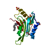
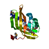
- PDB-3qio: Crystal Structure of HIV-1 RNase H with engineered E. coli loop a... -
+ Open data

- Basic information
| Entry | Database: PDB / ID: 3qio | ||||||
|---|---|---|---|---|---|---|---|
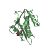
| Title | Crystal Structure of HIV-1 RNase H with engineered E. coli loop and N-hydroxy quinazolinedione inhibitor | ||||||
 Components Components | Gag-Pol polyprotein,Ribonuclease HI,Gag-Pol polyprotein | ||||||
 Keywords Keywords | TRANSFERASE / HYDROLASE/INHIBITOR / RNase H / HIV-1 / inhibitor / nuclease / HYDROLASE-INHIBITOR complex | ||||||
| Function / homology |  Function and homology information Function and homology informationDNA replication, removal of RNA primer / integrase activity / Integration of viral DNA into host genomic DNA / Autointegration results in viral DNA circles / ribonuclease H / Minus-strand DNA synthesis / Plus-strand DNA synthesis / Uncoating of the HIV Virion / 2-LTR circle formation / Vpr-mediated nuclear import of PICs ...DNA replication, removal of RNA primer / integrase activity / Integration of viral DNA into host genomic DNA / Autointegration results in viral DNA circles / ribonuclease H / Minus-strand DNA synthesis / Plus-strand DNA synthesis / Uncoating of the HIV Virion / 2-LTR circle formation / Vpr-mediated nuclear import of PICs / Early Phase of HIV Life Cycle / Integration of provirus / APOBEC3G mediated resistance to HIV-1 infection / Binding and entry of HIV virion / viral life cycle / HIV-1 retropepsin / symbiont-mediated activation of host apoptosis / retroviral ribonuclease H / exoribonuclease H / exoribonuclease H activity / Assembly Of The HIV Virion / protein processing / viral genome integration into host DNA / Budding and maturation of HIV virion / establishment of integrated proviral latency / RNA-directed DNA polymerase / RNA stem-loop binding / viral penetration into host nucleus / host multivesicular body / RNA-directed DNA polymerase activity / RNA-DNA hybrid ribonuclease activity / Transferases; Transferring phosphorus-containing groups; Nucleotidyltransferases / peptidase activity / host cell / viral nucleocapsid / endonuclease activity / DNA recombination / DNA-directed DNA polymerase / aspartic-type endopeptidase activity / Hydrolases; Acting on ester bonds / nucleic acid binding / DNA-directed DNA polymerase activity / symbiont-mediated suppression of host gene expression / viral translational frameshifting / symbiont entry into host cell / lipid binding / host cell nucleus / host cell plasma membrane / virion membrane / structural molecule activity / magnesium ion binding / DNA binding / zinc ion binding / identical protein binding / cytoplasm Similarity search - Function | ||||||
| Biological species |  Human immunodeficiency virus type 1 group M subtype B Human immunodeficiency virus type 1 group M subtype B | ||||||
| Method |  X-RAY DIFFRACTION / SYNCHROTRON / MOLECULAR REPLACEMENT / Resolution: 1.4011 Å X-RAY DIFFRACTION / SYNCHROTRON / MOLECULAR REPLACEMENT / Resolution: 1.4011 Å | ||||||
 Authors Authors | Lansdon, E.B. / Liu, Q. | ||||||
 Citation Citation | Journal: Antimicrob.Agents Chemother. / Year: 2011 Title: Structural and Binding Analysis of Pyrimidinol Carboxylic Acid and N-Hydroxy Quinazolinedione HIV-1 RNase H Inhibitors. Authors: Lansdon, E.B. / Liu, Q. / Leavitt, S.A. / Balakrishnan, M. / Perry, J.K. / Lancaster-Moyer, C. / Kutty, N. / Liu, X. / Squires, N.H. / Watkins, W.J. / Kirschberg, T.A. | ||||||
| History |
|
- Structure visualization
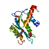
| Structure viewer | Molecule: MolmilJmol/JSmol |
|---|
- Downloads & links
-Download
| PDBx/mmCIF format | 3qio.cif.gz | 48.1 KB | Display | PDBx/mmCIF format |
|---|---|---|---|---|
| PDB format | pdb3qio.ent.gz | 31.6 KB | Display | PDB format |
| PDBx/mmJSON format | 3qio.json.gz | Tree view | PDBx/mmJSON format | |
| Others |  Other downloads Other downloads |
-Validation report
| Arichive directory | https://data.pdbj.org/pub/pdb/validation_reports/qi/3qioftp://data.pdbj.org/pub/pdb/validation_reports/qi/3qio | HTTPS FTP |
|---|
-Related structure data
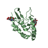
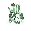
| Related structure data | 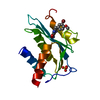 3qinC  3qipC  1hrhS S: Starting model for refinement C: citing same article ( |
|---|---|
| Similar structure data |


-Links
 PDBj
PDBj


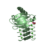
- Assembly
| Deposited unit | 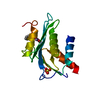
| ||||||||
|---|---|---|---|---|---|---|---|---|---|
| 1 |
| ||||||||
| Unit cell |
|
-Components
| #1: Protein | Mass: 16921.463 Da / Num. of mol.: 1 Fragment: HIV-1 RNase H (UNP REsdieus 1014-1148),HIV-1 RNase H (UNP REsdieus 1014-1148),HIV-1 RNase H (UNP REsdieus 1014-1148) Source method: isolated from a genetically manipulated source Source: (gene. exp.) Human immunodeficiency virus type 1 group M subtype B, (gene. exp.) Human immunodeficiency virus type 1 group M subtype B (isolate HXB2)Strain: isolate HXB2, K12 / Gene: gag-pol, rnhA, dasF, herA, rnh, sdrA, b0214, JW0204 / Plasmid: pET 30B / Production host: References: UniProt: P04585, UniProt: P0A7Y4, HIV-1 retropepsin, RNA-directed DNA polymerase, DNA-directed DNA polymerase, retroviral ribonuclease H, exoribonuclease H, Transferases; Transferring ...References: UniProt: P04585, UniProt: P0A7Y4, HIV-1 retropepsin, RNA-directed DNA polymerase, DNA-directed DNA polymerase, retroviral ribonuclease H, exoribonuclease H, Transferases; Transferring phosphorus-containing groups; Nucleotidyltransferases, Hydrolases; Acting on ester bonds, ribonuclease H | ||||||
|---|---|---|---|---|---|---|---|
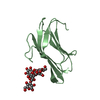
| #2: Chemical |   Mass: 54.938 Da / Num. of mol.: 2 / Source method: obtained synthetically / Formula: Mn Mass: 54.938 Da / Num. of mol.: 2 / Source method: obtained synthetically / Formula: Mn#3: Chemical | ChemComp-SO4 / |   Mass: 96.063 Da / Num. of mol.: 1 / Source method: obtained synthetically / Formula: SO4 Mass: 96.063 Da / Num. of mol.: 1 / Source method: obtained synthetically / Formula: SO4#4: Chemical | ChemComp-QID / | 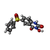  Mass: 318.305 Da / Num. of mol.: 1 / Source method: obtained synthetically / Formula: C14H10N2O5S Mass: 318.305 Da / Num. of mol.: 1 / Source method: obtained synthetically / Formula: C14H10N2O5S#5: Water | ChemComp-HOH / |  Mass: 18.015 Da / Num. of mol.: 185 / Source method: isolated from a natural source / Formula: H2O Mass: 18.015 Da / Num. of mol.: 185 / Source method: isolated from a natural source / Formula: H2O |
-Experimental details
-Experiment
| Experiment | Method: X-RAY DIFFRACTION / Number of used crystals: 1 |
|---|
- Sample preparation
| Crystal | Density Matthews: 2.29 Å3/Da / Density % sol: 46.23 % |
|---|---|
| Crystal grow | Temperature: 293 K / Method: vapor diffusion, hanging drop / pH: 7.5 Details: 15% PEG 3350, 100mM HEPES pH 7.5, and 200mM LiSO4, VAPOR DIFFUSION, HANGING DROP, temperature 293K |
-Data collection
| Diffraction | Mean temperature: 100 K |
|---|---|
| Diffraction source | Source: SYNCHROTRON / Site: ALS  / Beamline: 5.0.1 / Wavelength: 0.98 Å / Beamline: 5.0.1 / Wavelength: 0.98 Å |
| Detector | Type: ADSC QUANTUM 210 / Detector: CCD / Date: Oct 25, 2007 |
| Radiation | Monochromator: single crystal, cylindrically bent, SI(220) / Protocol: SINGLE WAVELENGTH / Monochromatic (M) / Laue (L): M / Scattering type: x-ray |
| Radiation wavelength | Wavelength: 0.98 Å / Relative weight: 1 |
| Reflection | Resolution: 1.4→50 Å / Num. obs: 30610 / % possible obs: 98 % / Observed criterion σ(F): 0 / Observed criterion σ(I): 0 / Redundancy: 5.3 % / Rmerge(I) obs: 0.06 / Net I/σ(I): 19.3 |
| Reflection shell | Resolution: 1.4→1.43 Å / Redundancy: 4 % / Rmerge(I) obs: 0.497 / Mean I/σ(I) obs: 1.7 / % possible all: 83.1 |
- Processing
| Software |
| |||||||||||||||||||||||||||||||||||||||||||||||||||||||||||||||||||||||||||||||||||||||||||||||||||||||||
|---|---|---|---|---|---|---|---|---|---|---|---|---|---|---|---|---|---|---|---|---|---|---|---|---|---|---|---|---|---|---|---|---|---|---|---|---|---|---|---|---|---|---|---|---|---|---|---|---|---|---|---|---|---|---|---|---|---|---|---|---|---|---|---|---|---|---|---|---|---|---|---|---|---|---|---|---|---|---|---|---|---|---|---|---|---|---|---|---|---|---|---|---|---|---|---|---|---|---|---|---|---|---|---|---|---|---|
| Refinement | Method to determine structure: MOLECULAR REPLACEMENT Starting model: PDB ENTRY 1HRH Resolution: 1.4011→28.176 Å / SU ML: 0.19 / Cross valid method: THROUGHOUT / σ(F): 0 / Phase error: 21.91 / Stereochemistry target values: ML
| |||||||||||||||||||||||||||||||||||||||||||||||||||||||||||||||||||||||||||||||||||||||||||||||||||||||||
| Solvent computation | Shrinkage radii: 1.06 Å / VDW probe radii: 1.3 Å / Solvent model: FLAT BULK SOLVENT MODEL / Bsol: 45.216 Å2 / ksol: 0.332 e/Å3 | |||||||||||||||||||||||||||||||||||||||||||||||||||||||||||||||||||||||||||||||||||||||||||||||||||||||||
| Displacement parameters |
| |||||||||||||||||||||||||||||||||||||||||||||||||||||||||||||||||||||||||||||||||||||||||||||||||||||||||
| Refinement step | Cycle: LAST / Resolution: 1.4011→28.176 Å
| |||||||||||||||||||||||||||||||||||||||||||||||||||||||||||||||||||||||||||||||||||||||||||||||||||||||||
| Refine LS restraints |
| |||||||||||||||||||||||||||||||||||||||||||||||||||||||||||||||||||||||||||||||||||||||||||||||||||||||||
| LS refinement shell |
|
