 Movie
Movie Controller

[English] 日本語
 Yorodumi
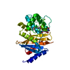
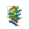
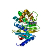
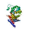
- PDB-3pag: Crystal structure of the V130D mutant of OXA-24/40 in complex wit... -
+ Open data

- Basic information
| Entry | Database: PDB / ID: 3pag | ||||||
|---|---|---|---|---|---|---|---|
| Title | Crystal structure of the V130D mutant of OXA-24/40 in complex with doripenem | ||||||
 Components Components | Beta-lactamase | ||||||
 Keywords Keywords | HYDROLASE/ANTIBIOTIC / hydrolase / carbapenemase / HYDROLASE-ANTIBIOTIC complex | ||||||
| Function / homology |  Function and homology information Function and homology information | ||||||
| Biological species |  Acinetobacter baumannii (bacteria) Acinetobacter baumannii (bacteria) | ||||||
| Method |  X-RAY DIFFRACTION / SYNCHROTRON / MOLECULAR REPLACEMENT / Resolution: 2.25 Å X-RAY DIFFRACTION / SYNCHROTRON / MOLECULAR REPLACEMENT / Resolution: 2.25 Å | ||||||
 Authors Authors | Powers, R.A. / Leonard, D.A. / Schneider, K.D. | ||||||
 Citation Citation | Journal: J.Mol.Biol. / Year: 2011 Title: Structures of the Class D Carbapenemase OXA-24 from Acinetobacter baumannii in Complex with Doripenem. Authors: Schneider, K.D. / Ortega, C.J. / Renck, N.A. / Bonomo, R.A. / Powers, R.A. / Leonard, D.A. #1: Journal: Biochemistry / Year: 2009Title: The 1.4 A crystal structure of the class D beta-lactamase OXA-1 complexed with doripenem Authors: Schneider, K.D. / Karpen, M.E. / Bonomo, R.A. / Leonard, D.A. / Powers, R.A. | ||||||
| History |
|
- Structure visualization
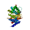
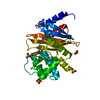
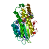
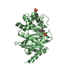
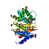
| Structure viewer | Molecule: MolmilJmol/JSmol |
|---|
- Downloads & links
-Download
| PDBx/mmCIF format | 3pag.cif.gz | 114.9 KB | Display | PDBx/mmCIF format |
|---|---|---|---|---|
| PDB format | pdb3pag.ent.gz | 89 KB | Display | PDB format |
| PDBx/mmJSON format | 3pag.json.gz | Tree view | PDBx/mmJSON format | |
| Others |  Other downloads Other downloads |
-Validation report
| Arichive directory | https://data.pdbj.org/pub/pdb/validation_reports/pa/3pagftp://data.pdbj.org/pub/pdb/validation_reports/pa/3pag | HTTPS FTP |
|---|
-Related structure data
| Related structure data | 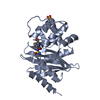 3paeC 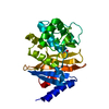 2jc7S C: citing same article ( S: Starting model for refinement |
|---|---|
| Similar structure data |


-Links
 PDBj
PDBj


- Assembly
| Deposited unit | 
| ||||||||
|---|---|---|---|---|---|---|---|---|---|
| 1 | 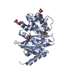
| ||||||||
| 2 | 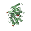
| ||||||||
| Unit cell |
|
-Components
| #1: Protein | Mass: 27690.809 Da / Num. of mol.: 2 / Fragment: UNP residues 32-275 / Mutation: V130D Source method: isolated from a genetically manipulated source Source: (gene. exp.) Acinetobacter baumannii (bacteria)Gene: blaOXA-33, bla-OXA-40, blaOXA-24, blaOXA-40, oxa-24, oxa40 Plasmid: pET24a / Production host: #2: Chemical | 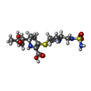  Mass: 422.520 Da / Num. of mol.: 2 / Source method: obtained synthetically / Formula: C15H26N4O6S2 Mass: 422.520 Da / Num. of mol.: 2 / Source method: obtained synthetically / Formula: C15H26N4O6S2#3: Chemical | ChemComp-SO4 /   Mass: 96.063 Da / Num. of mol.: 6 / Source method: obtained synthetically / Formula: SO4 Mass: 96.063 Da / Num. of mol.: 6 / Source method: obtained synthetically / Formula: SO4#4: Water | ChemComp-HOH / |  Mass: 18.015 Da / Num. of mol.: 196 / Source method: isolated from a natural source / Formula: H2O Mass: 18.015 Da / Num. of mol.: 196 / Source method: isolated from a natural source / Formula: H2OHas protein modification | Y | Nonpolymer details | 4J6 IN THE STRUCTURE REPRESENTS THE DELTA 2 TAUTOMER, IN WHICH A DOUBLE BOND IS PRESENT BETWEEN ...4J6 IN THE STRUCTURE REPRESENTS | |
|---|
-Experimental details
-Experiment
| Experiment | Method: X-RAY DIFFRACTION / Number of used crystals: 1 |
|---|
- Sample preparation
| Crystal | Density Matthews: 4.03 Å3/Da / Density % sol: 69.51 % |
|---|---|
| Crystal grow | Temperature: 298 K / Method: vapor diffusion, hanging drop / pH: 8.5 Details: 100 mM TRIS-HCl, 2.0 M ammonium sulfate, pH 8.5, VAPOR DIFFUSION, HANGING DROP, temperature 298K |
-Data collection
| Diffraction | Mean temperature: 100 K |
|---|---|
| Diffraction source | Source: SYNCHROTRON / Site: APS  / Beamline: 21-ID-D / Wavelength: 1.1 Å / Beamline: 21-ID-D / Wavelength: 1.1 Å |
| Detector | Type: MARMOSAIC 300 mm CCD / Detector: CCD / Date: Jun 5, 2009 |
| Radiation | Monochromator: Si(111) / Protocol: SINGLE WAVELENGTH / Monochromatic (M) / Laue (L): M / Scattering type: x-ray |
| Radiation wavelength | Wavelength: 1.1 Å / Relative weight: 1 |
| Reflection | Resolution: 2.25→50 Å / Num. obs: 41616 / % possible obs: 99 % |
| Reflection shell | Resolution: 2.25→2.33 Å / % possible all: 95.5 |
- Processing
| Software |
| ||||||||||||||||||||||||||||||||||||||||||||||||||||||||||||||||||||||||||||||||||||||||||||||||||||||||||||||||||||||||||||||||||||||||||||||||||||||||||||||||||||||||||
|---|---|---|---|---|---|---|---|---|---|---|---|---|---|---|---|---|---|---|---|---|---|---|---|---|---|---|---|---|---|---|---|---|---|---|---|---|---|---|---|---|---|---|---|---|---|---|---|---|---|---|---|---|---|---|---|---|---|---|---|---|---|---|---|---|---|---|---|---|---|---|---|---|---|---|---|---|---|---|---|---|---|---|---|---|---|---|---|---|---|---|---|---|---|---|---|---|---|---|---|---|---|---|---|---|---|---|---|---|---|---|---|---|---|---|---|---|---|---|---|---|---|---|---|---|---|---|---|---|---|---|---|---|---|---|---|---|---|---|---|---|---|---|---|---|---|---|---|---|---|---|---|---|---|---|---|---|---|---|---|---|---|---|---|---|---|---|---|---|---|---|---|
| Refinement | Method to determine structure: MOLECULAR REPLACEMENT Starting model: 2JC7 Resolution: 2.25→50 Å / Cor.coef. Fo:Fc: 0.956 / Cor.coef. Fo:Fc free: 0.935 / SU B: 4.934 / SU ML: 0.122 / Cross valid method: THROUGHOUT / ESU R: 0.186 / ESU R Free: 0.174 / Stereochemistry target values: MAXIMUM LIKELIHOOD / Details: HYDROGENS HAVE BEEN ADDED IN THE RIDING POSITIONS
| ||||||||||||||||||||||||||||||||||||||||||||||||||||||||||||||||||||||||||||||||||||||||||||||||||||||||||||||||||||||||||||||||||||||||||||||||||||||||||||||||||||||||||
| Solvent computation | Ion probe radii: 0.8 Å / Shrinkage radii: 0.8 Å / VDW probe radii: 1.4 Å / Solvent model: BABINET MODEL WITH MASK | ||||||||||||||||||||||||||||||||||||||||||||||||||||||||||||||||||||||||||||||||||||||||||||||||||||||||||||||||||||||||||||||||||||||||||||||||||||||||||||||||||||||||||
| Displacement parameters | Biso mean: 36.791 Å2
| ||||||||||||||||||||||||||||||||||||||||||||||||||||||||||||||||||||||||||||||||||||||||||||||||||||||||||||||||||||||||||||||||||||||||||||||||||||||||||||||||||||||||||
| Refinement step | Cycle: LAST / Resolution: 2.25→50 Å
| ||||||||||||||||||||||||||||||||||||||||||||||||||||||||||||||||||||||||||||||||||||||||||||||||||||||||||||||||||||||||||||||||||||||||||||||||||||||||||||||||||||||||||
| Refine LS restraints |
| ||||||||||||||||||||||||||||||||||||||||||||||||||||||||||||||||||||||||||||||||||||||||||||||||||||||||||||||||||||||||||||||||||||||||||||||||||||||||||||||||||||||||||
| LS refinement shell | Resolution: 2.248→2.306 Å / Total num. of bins used: 20
|
