 Movie
Movie Controller

[English] 日本語
 Yorodumi
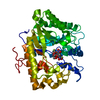
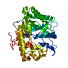
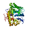
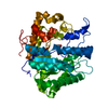
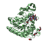
- PDB-3n9k: F229A/E292S Double Mutant of Exo-beta-1,3-glucanase from Candida ... -
+ Open data

- Basic information
| Entry | Database: PDB / ID: 3n9k | |||||||||
|---|---|---|---|---|---|---|---|---|---|---|
| Title | F229A/E292S Double Mutant of Exo-beta-1,3-glucanase from Candida albicans in Complex with Laminaritriose at 1.7 A | |||||||||
 Components Components | Glucan 1,3-beta-glucosidase | |||||||||
 Keywords Keywords | HYDROLASE / Aromatic entranceway/clamp / Exoglucanase / Glycoside hydrolase / Protein-carbohydrate interaction / Site-directed mutagenesis / Laminaritriose | |||||||||
| Function / homology |  Function and homology information Function and homology informationsingle-species biofilm formation in or on host organism / glucan metabolic process / glucan 1,3-beta-glucosidase / single-species biofilm formation on inanimate substrate / glucan exo-1,3-beta-glucosidase activity / adhesion of symbiont to host cell / fungal-type cell wall organization / glucan catabolic process / Transferases; Glycosyltransferases; Hexosyltransferases / cell-substrate adhesion ...single-species biofilm formation in or on host organism / glucan metabolic process / glucan 1,3-beta-glucosidase / single-species biofilm formation on inanimate substrate / glucan exo-1,3-beta-glucosidase activity / adhesion of symbiont to host cell / fungal-type cell wall organization / glucan catabolic process / Transferases; Glycosyltransferases; Hexosyltransferases / cell-substrate adhesion / cell adhesion molecule binding / extracellular vesicle / transferase activity / cell surface / extracellular region Similarity search - Function | |||||||||
| Biological species |  Candida albicans (yeast) Candida albicans (yeast) | |||||||||
| Method |  X-RAY DIFFRACTION / MOLECULAR REPLACEMENT / Resolution: 1.7 Å X-RAY DIFFRACTION / MOLECULAR REPLACEMENT / Resolution: 1.7 Å | |||||||||
 Authors Authors | Nakatani, Y. / Cutfield, S.M. / Cutfield, J.F. | |||||||||
 Citation Citation | Journal: Febs J. / Year: 2010 Title: Carbohydrate binding sites in Candida albicans exo-beta-1,3-glucanase and the role of the Phe-Phe 'clamp' at the active site entrance Authors: Patrick, W.M. / Nakatani, Y. / Cutfield, S.M. / Sharpe, M.L. / Ramsay, R.J. / Cutfield, J.F. | |||||||||
| History |
|
- Structure visualization
| Structure viewer | Molecule: MolmilJmol/JSmol |
|---|
- Downloads & links
-Download
| PDBx/mmCIF format | 3n9k.cif.gz | 105.5 KB | Display | PDBx/mmCIF format |
|---|---|---|---|---|
| PDB format | pdb3n9k.ent.gz | 78.9 KB | Display | PDB format |
| PDBx/mmJSON format | 3n9k.json.gz | Tree view | PDBx/mmJSON format | |
| Others |  Other downloads Other downloads |
-Validation report
| Arichive directory | https://data.pdbj.org/pub/pdb/validation_reports/n9/3n9kftp://data.pdbj.org/pub/pdb/validation_reports/n9/3n9k | HTTPS FTP |
|---|
-Related structure data
| Related structure data | 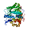 2pc8C  2pf0C 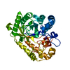 3o6aC 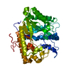 1cz1S S: Starting model for refinement C: citing same article ( |
|---|---|
| Similar structure data |


-Links
 PDBj
PDBj- Assembly
| Deposited unit | 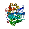
| ||||||||
|---|---|---|---|---|---|---|---|---|---|
| 1 |
| ||||||||
| Unit cell |
|
-Components
| #1: Protein | Mass: 45617.219 Da / Num. of mol.: 1 / Mutation: F229A,E292S Source method: isolated from a genetically manipulated source Source: (gene. exp.) Candida albicans (yeast) / Strain: ATCC 10261 / Gene: XOG / Plasmid: PPIC9K / Production host:  Pichia pastoris (fungus) / Strain (production host): KM71 / References: UniProt: P29717, glucan 1,3-beta-glucosidase Pichia pastoris (fungus) / Strain (production host): KM71 / References: UniProt: P29717, glucan 1,3-beta-glucosidase |
|---|---|
| #2: Polysaccharide | beta-D-glucopyranose-(1-3)-beta-D-glucopyranose-(1-3)-beta-D-glucopyranose Source method: isolated from a genetically manipulated source |
| #3: Polysaccharide | beta-D-glucopyranose-(1-3)-beta-D-glucopyranose / beta-laminaribiose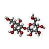  Source method: isolated from a genetically manipulated source Details: oligosaccharide / References: beta-laminaribiose |
| #4: Chemical | ChemComp-CA /   Mass: 40.078 Da / Num. of mol.: 1 / Source method: obtained synthetically / Formula: Ca Mass: 40.078 Da / Num. of mol.: 1 / Source method: obtained synthetically / Formula: Ca |
| #5: Water | ChemComp-HOH /  Mass: 18.015 Da / Num. of mol.: 404 / Source method: isolated from a natural source / Formula: H2O Mass: 18.015 Da / Num. of mol.: 404 / Source method: isolated from a natural source / Formula: H2O |
| Has protein modification | Y |
| Sequence details | DUE TO ALTERNATIVE CODON USAGE BY CANDIDA ALBICANS, THIS POSITION IS A SER WHEN FROM NATURAL ...DUE TO ALTERNATIV |
-Experimental details
-Experiment
| Experiment | Method: X-RAY DIFFRACTION / Number of used crystals: 1 |
|---|
- Sample preparation
| Crystal | Density Matthews: 1.97 Å3/Da / Density % sol: 37.44 % |
|---|---|
| Crystal grow | Temperature: 291 K / Method: vapor diffusion, hanging drop / pH: 7.3 Details: 0.1M HEPES-KOH, 0.2M CACL2, 19% PEG 8000, pH 7.3, VAPOR DIFFUSION, HANGING DROP, temperature 291K |
-Data collection
| Diffraction | Mean temperature: 113 K |
|---|---|
| Diffraction source | Source: ROTATING ANODE / Type: RIGAKU MICROMAX-007 HF / Wavelength: 1.5418 Å |
| Detector | Type: RIGAKU RAXIS IV / Detector: IMAGE PLATE / Date: Mar 10, 2010 |
| Radiation | Protocol: SINGLE WAVELENGTH / Monochromatic (M) / Laue (L): M / Scattering type: x-ray |
| Radiation wavelength | Wavelength: 1.5418 Å / Relative weight: 1 |
| Reflection | Resolution: 1.7→38.19 Å / Num. all: 40339 / Num. obs: 40247 / % possible obs: 99.8 % / Redundancy: 4.2 % / Biso Wilson estimate: 17.8 Å2 / Rmerge(I) obs: 0.074 / Rsym value: 0.074 / Net I/σ(I): 14.1 |
| Reflection shell | Resolution: 1.7→1.79 Å / Redundancy: 4.1 % / Rmerge(I) obs: 0.636 / Mean I/σ(I) obs: 2.2 / Num. unique all: 5803 / Rsym value: 0.636 / % possible all: 99.9 |
- Processing
| Software |
| |||||||||||||||||||||||||||||||||||||||||||||||||||||||||||||||||
|---|---|---|---|---|---|---|---|---|---|---|---|---|---|---|---|---|---|---|---|---|---|---|---|---|---|---|---|---|---|---|---|---|---|---|---|---|---|---|---|---|---|---|---|---|---|---|---|---|---|---|---|---|---|---|---|---|---|---|---|---|---|---|---|---|---|---|
| Refinement | Method to determine structure: MOLECULAR REPLACEMENT Starting model: PDB ENTRY 1CZ1 Resolution: 1.7→38.19 Å / Cor.coef. Fo:Fc: 0.97 / Cor.coef. Fo:Fc free: 0.954 / SU B: 2.077 / SU ML: 0.068 / Isotropic thermal model: Isotropic / Cross valid method: THROUGHOUT / ESU R Free: 0.106 / Stereochemistry target values: MAXIMUM LIKELIHOOD / Details: HYDROGENS HAVE BEEN ADDED IN THE RIDING POSITIONS
| |||||||||||||||||||||||||||||||||||||||||||||||||||||||||||||||||
| Solvent computation | Ion probe radii: 0.8 Å / Shrinkage radii: 0.8 Å / VDW probe radii: 1.4 Å / Solvent model: MASK | |||||||||||||||||||||||||||||||||||||||||||||||||||||||||||||||||
| Displacement parameters | Biso mean: 17.546 Å2
| |||||||||||||||||||||||||||||||||||||||||||||||||||||||||||||||||
| Refinement step | Cycle: LAST / Resolution: 1.7→38.19 Å
| |||||||||||||||||||||||||||||||||||||||||||||||||||||||||||||||||
| Refine LS restraints |
| |||||||||||||||||||||||||||||||||||||||||||||||||||||||||||||||||
| LS refinement shell | Resolution: 1.7→1.744 Å / Total num. of bins used: 20
|
