 Movie
Movie Controller

[English] 日本語
 Yorodumi
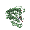
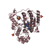
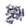
- PDB-4m81: The structure of E292S glycosynthase variant of exo-1,3-beta-gluc... -
+ Open data

- Basic information
| Entry | Database: PDB / ID: 4m81 | ||||||
|---|---|---|---|---|---|---|---|
| Title | The structure of E292S glycosynthase variant of exo-1,3-beta-glucanase from Candida albicans complexed with 1-fluoro-alpha-D-glucopyranoside (donor) and p-nitrophenyl beta-D-glucopyranoside (acceptor) at 1.86A resolution | ||||||
 Components Components | EXO-1,3-BETA-GLUCANASE | ||||||
 Keywords Keywords | HYDROLASE / TIM BARREL / GLYCOSIDE HYDROLASE FAMILY 5 / GLYCOSIDE HYDROLASE / CELL WALL HYDROLASE / GLYCOSYNTHASE / PROTEIN-CARBOHYDRATE INTERACTION | ||||||
| Function / homology |  Function and homology information Function and homology informationsingle-species biofilm formation in or on host organism / fungal-type cell wall (1->3)-beta-D-glucan metabolic process / glucan metabolic process / glucan 1,3-beta-glucosidase / single-species biofilm formation on inanimate substrate / glucan exo-1,3-beta-glucosidase activity / adhesion of symbiont to host cell / fungal-type cell wall organization / glucan catabolic process / Transferases; Glycosyltransferases; Hexosyltransferases ...single-species biofilm formation in or on host organism / fungal-type cell wall (1->3)-beta-D-glucan metabolic process / glucan metabolic process / glucan 1,3-beta-glucosidase / single-species biofilm formation on inanimate substrate / glucan exo-1,3-beta-glucosidase activity / adhesion of symbiont to host cell / fungal-type cell wall organization / glucan catabolic process / Transferases; Glycosyltransferases; Hexosyltransferases / cell-substrate adhesion / cell adhesion molecule binding / extracellular vesicle / transferase activity / cell surface / extracellular region Similarity search - Function | ||||||
| Biological species |  Candida albicans (yeast) Candida albicans (yeast) | ||||||
| Method |  X-RAY DIFFRACTION / MOLECULAR REPLACEMENT / Resolution: 1.86 Å X-RAY DIFFRACTION / MOLECULAR REPLACEMENT / Resolution: 1.86 Å | ||||||
 Authors Authors | Nakatani, Y. / Cutfield, S.M. / Larsen, D.S. / Cutfield, J.F. | ||||||
 Citation Citation | Journal: Biochemistry / Year: 2014 Title: Major Change in Regiospecificity for the Exo-1,3-beta-glucanase from Candida albicans following Its Conversion to a Glycosynthase. Authors: Nakatani, Y. / Larsen, D.S. / Cutfield, S.M. / Cutfield, J.F. | ||||||
| History |
|
- Structure visualization
| Structure viewer | Molecule: MolmilJmol/JSmol |
|---|
- Downloads & links
-Download
| PDBx/mmCIF format | 4m81.cif.gz | 106.6 KB | Display | PDBx/mmCIF format |
|---|---|---|---|---|
| PDB format | pdb4m81.ent.gz | 80.2 KB | Display | PDB format |
| PDBx/mmJSON format | 4m81.json.gz | Tree view | PDBx/mmJSON format | |
| Others |  Other downloads Other downloads |
-Validation report
| Arichive directory | https://data.pdbj.org/pub/pdb/validation_reports/m8/4m81ftp://data.pdbj.org/pub/pdb/validation_reports/m8/4m81 | HTTPS FTP |
|---|
-Related structure data
| Related structure data |  4m80C  4m82C  1cz1S S: Starting model for refinement C: citing same article ( |
|---|---|
| Similar structure data |


-Links
 PDBj
PDBj- Assembly
| Deposited unit | 
| ||||||||
|---|---|---|---|---|---|---|---|---|---|
| 1 |
| ||||||||
| Unit cell |
|
-Components
| #1: Protein | Mass: 45693.312 Da / Num. of mol.: 1 / Fragment: EXO-1,3-BETA-GLUCANASE (unp residues 40-438) / Mutation: E292S Source method: isolated from a genetically manipulated source Source: (gene. exp.) Candida albicans (yeast) / Strain: ATCC 10261 / Gene: CaO19.10507, XOG, XOG1 / Plasmid: PPIC9K / Production host:  Komagataella pastoris (fungus) / Strain (production host): GS115/KM71 Komagataella pastoris (fungus) / Strain (production host): GS115/KM71References: UniProt: Q5AI63, UniProt: P29717*PLUS, glucan 1,3-beta-glucosidase |
|---|---|
| #2: Sugar | ChemComp-GLF /   Type: D-saccharide / Mass: 182.147 Da / Num. of mol.: 1 Type: D-saccharide / Mass: 182.147 Da / Num. of mol.: 1Source method: isolated from a genetically manipulated source Formula: C6H11FO5 |
| #3: Sugar | ChemComp-PNW / 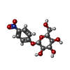  Type: D-saccharide / Mass: 301.249 Da / Num. of mol.: 1 Type: D-saccharide / Mass: 301.249 Da / Num. of mol.: 1Source method: isolated from a genetically manipulated source Formula: C12H15NO8 |
| #4: Chemical | ChemComp-GOL /   Mass: 92.094 Da / Num. of mol.: 1 / Source method: obtained synthetically / Formula: C3H8O3 Mass: 92.094 Da / Num. of mol.: 1 / Source method: obtained synthetically / Formula: C3H8O3 |
| #5: Water | ChemComp-HOH /  Mass: 18.015 Da / Num. of mol.: 492 / Source method: isolated from a natural source / Formula: H2O Mass: 18.015 Da / Num. of mol.: 492 / Source method: isolated from a natural source / Formula: H2O |
| Has protein modification | Y |
| Sequence details | DUE TO ALTERNATIVE CODON USAGE BY CANDIDA ALBICANS, THIS POSITION IS A SER WHEN FROM NATURAL ...DUE TO ALTERNATIV |
-Experimental details
-Experiment
| Experiment | Method: X-RAY DIFFRACTION / Number of used crystals: 1 |
|---|
- Sample preparation
| Crystal | Density Matthews: 1.97 Å3/Da / Density % sol: 37.56 % |
|---|---|
| Crystal grow | Temperature: 291 K / Method: vapor diffusion, hanging drop / pH: 7.3 Details: 0.1M HEPES-KOH, 0.2M CACL2, 19% PEG8000, pH 7.3, VAPOR DIFFUSION, HANGING DROP, temperature 291K |
-Data collection
| Diffraction | Mean temperature: 113 K |
|---|---|
| Diffraction source | Source: ROTATING ANODE / Type: RIGAKU MICROMAX-007 HF / Wavelength: 1.5418 Å |
| Detector | Type: MAR scanner 345 mm plate / Detector: IMAGE PLATE / Date: Jun 6, 2004 |
| Radiation | Protocol: SINGLE WAVELENGTH / Monochromatic (M) / Laue (L): M / Scattering type: x-ray |
| Radiation wavelength | Wavelength: 1.5418 Å / Relative weight: 1 |
| Reflection | Resolution: 1.86→37.01 Å / Num. all: 28317 / Num. obs: 28249 / % possible obs: 90.7 % / Redundancy: 5.2 % / Biso Wilson estimate: 17.2 Å2 / Rmerge(I) obs: 0.088 / Rsym value: 0.088 / Net I/σ(I): 15.5 |
| Reflection shell | Resolution: 1.86→1.96 Å / Redundancy: 3.9 % / Rmerge(I) obs: 0.527 / Mean I/σ(I) obs: 2.7 / Num. unique all: 3304 / Rsym value: 0.527 / % possible all: 74 |
- Processing
| Software |
| |||||||||||||||||||||||||||||||||||||||||||||||||||||||||||||||||||||||||||||
|---|---|---|---|---|---|---|---|---|---|---|---|---|---|---|---|---|---|---|---|---|---|---|---|---|---|---|---|---|---|---|---|---|---|---|---|---|---|---|---|---|---|---|---|---|---|---|---|---|---|---|---|---|---|---|---|---|---|---|---|---|---|---|---|---|---|---|---|---|---|---|---|---|---|---|---|---|---|---|
| Refinement | Method to determine structure: MOLECULAR REPLACEMENT Starting model: pdb entry 1CZ1 Resolution: 1.86→18.092 Å / SU ML: 0.2 / Cross valid method: THROUGHOUT / σ(F): 1.34 / Phase error: 20.09 / Stereochemistry target values: ML
| |||||||||||||||||||||||||||||||||||||||||||||||||||||||||||||||||||||||||||||
| Solvent computation | Shrinkage radii: 0.9 Å / VDW probe radii: 1.11 Å / Solvent model: FLAT BULK SOLVENT MODEL | |||||||||||||||||||||||||||||||||||||||||||||||||||||||||||||||||||||||||||||
| Refinement step | Cycle: LAST / Resolution: 1.86→18.092 Å
| |||||||||||||||||||||||||||||||||||||||||||||||||||||||||||||||||||||||||||||
| Refine LS restraints |
| |||||||||||||||||||||||||||||||||||||||||||||||||||||||||||||||||||||||||||||
| LS refinement shell |
|
