 Movie
Movie Controller


+ Open data

- Basic information
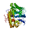
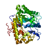
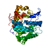
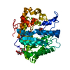
| Entry | Database: PDB / ID: 1eqp | ||||||
|---|---|---|---|---|---|---|---|
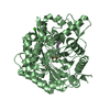
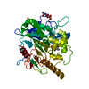
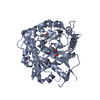
| Title | EXO-B-(1,3)-GLUCANASE FROM CANDIDA ALBICANS | ||||||
 Components Components | EXO-B-(1,3)-GLUCANASE | ||||||
 Keywords Keywords | HYDROLASE / CANDIDA ALBICANS / EXOGLUCANASE / ALTERNATIVE CODON USAGE | ||||||
| Function / homology |  Function and homology information Function and homology informationsingle-species biofilm formation in or on host organism / fungal-type cell wall (1->3)-beta-D-glucan metabolic process / glucan metabolic process / glucan 1,3-beta-glucosidase / single-species biofilm formation on inanimate substrate / glucan exo-1,3-beta-glucosidase activity / adhesion of symbiont to host cell / fungal-type cell wall organization / glucan catabolic process / Transferases; Glycosyltransferases; Hexosyltransferases ...single-species biofilm formation in or on host organism / fungal-type cell wall (1->3)-beta-D-glucan metabolic process / glucan metabolic process / glucan 1,3-beta-glucosidase / single-species biofilm formation on inanimate substrate / glucan exo-1,3-beta-glucosidase activity / adhesion of symbiont to host cell / fungal-type cell wall organization / glucan catabolic process / Transferases; Glycosyltransferases; Hexosyltransferases / cell-substrate adhesion / cell adhesion molecule binding / extracellular vesicle / transferase activity / cell surface / extracellular region Similarity search - Function | ||||||
| Biological species |  Candida albicans (yeast) Candida albicans (yeast) | ||||||
| Method |  X-RAY DIFFRACTION / Resolution: 1.9 Å X-RAY DIFFRACTION / Resolution: 1.9 Å | ||||||
 Authors Authors | Cutfield, J.F. / Sullivan, P.A. / Cutfield, S.M. | ||||||
 Citation Citation | Journal: Protein Eng. / Year: 2000 Title: Minor structural consequences of alternative CUG codon usage (Ser for Leu) in Candida albicans exoglucanase. Authors: Cutfield, J.F. / Sullivan, P.A. / Cutfield, S.M. #1: Journal: J.Mol.Biol. / Year: 1999Title: The Structure of the Exo-Beta-(1,3)-Glucanase from Candida albicans in Native and Bound Forms: Relationship Between a Pocket and Groove in Family 5 Glycosyl Hydrolases Authors: Cutfield, S.M. / Davies, G.J. / Murshudov, G. / Anderson, B.F. / Moody, P.C. / Sullivan, P.A. / Cutfield, J.F. | ||||||
| History |
|
- Structure visualization
| Structure viewer | Molecule: MolmilJmol/JSmol |
|---|
- Downloads & links
-Download
| PDBx/mmCIF format | 1eqp.cif.gz | 96.3 KB | Display | PDBx/mmCIF format |
|---|---|---|---|---|
| PDB format | pdb1eqp.ent.gz | 73.7 KB | Display | PDB format |
| PDBx/mmJSON format | 1eqp.json.gz | Tree view | PDBx/mmJSON format | |
| Others |  Other downloads Other downloads |
-Validation report
| Arichive directory | https://data.pdbj.org/pub/pdb/validation_reports/eq/1eqpftp://data.pdbj.org/pub/pdb/validation_reports/eq/1eqp | HTTPS FTP |
|---|
-Related structure data
| Related structure data | |
|---|---|
| Similar structure data |


-Links
 PDBj
PDBj- Assembly
| Deposited unit | 
| ||||||||
|---|---|---|---|---|---|---|---|---|---|
| 1 |
| ||||||||
| Unit cell |
|
-Components
| #1: Protein | Mass: 45243.797 Da / Num. of mol.: 1 / Source method: isolated from a natural source / Source: (natural) Candida albicans (yeast) / Strain: ATCC10261 / References: UniProt: P29717, glucan 1,3-beta-glucosidase |
|---|---|
| #2: Water | ChemComp-HOH /  Mass: 18.015 Da / Num. of mol.: 282 / Source method: isolated from a natural source / Formula: H2O Mass: 18.015 Da / Num. of mol.: 282 / Source method: isolated from a natural source / Formula: H2O |
| Has protein modification | Y |
-Experimental details
-Experiment
| Experiment | Method: X-RAY DIFFRACTION / Number of used crystals: 1 |
|---|
- Sample preparation
| Crystal | Density Matthews: 2.09 Å3/Da / Density % sol: 41.14 % |
|---|---|
| Crystal grow | Temperature: 291 K / Method: vapor diffusion, hanging drop / pH: 7.3 Details: HEPES BUFFER, CACL2, PEG 8000, pH 7.3, VAPOR DIFFUSION, HANGING DROP, temperature 291K |
| Crystal grow | *PLUS Details: Chambers, R.S., (1993) J. Gen. Microbiol., 139, 325., Chambers, R.S., (1993) FEBS Lett., 327, 366. |
-Data collection
| Diffraction | Mean temperature: 293 K |
|---|---|
| Diffraction source | Source: ROTATING ANODE / Type: RIGAKU RU200 / Wavelength: 1.5418 |
| Detector | Type: RIGAKU RAXIS II / Detector: IMAGE PLATE / Date: May 12, 1993 |
| Radiation | Protocol: SINGLE WAVELENGTH / Monochromatic (M) / Laue (L): M / Scattering type: x-ray |
| Radiation wavelength | Wavelength: 1.5418 Å / Relative weight: 1 |
| Reflection | Resolution: 1.93→54 Å / Num. all: 29591 / Num. obs: 26304 / % possible obs: 90.3 % / Observed criterion σ(F): 1 / Redundancy: 2.5 % / Biso Wilson estimate: 22.8 Å2 / Rmerge(I) obs: 0.035 / Net I/σ(I): 14.2 |
| Reflection shell | Resolution: 1.9→2.01 Å / Redundancy: 2.5 % / Rmerge(I) obs: 0.103 / % possible all: 48.7 |
| Reflection | *PLUS Highest resolution: 1.9 Å |
| Reflection shell | *PLUS % possible obs: 56.6 % / Mean I/σ(I) obs: 7 |
- Processing
| Software |
| ||||||||||||||||||||
|---|---|---|---|---|---|---|---|---|---|---|---|---|---|---|---|---|---|---|---|---|---|
| Refinement | Resolution: 1.9→20 Å / σ(F): 0 / Stereochemistry target values: Engh & Huber
| ||||||||||||||||||||
| Refinement step | Cycle: LAST / Resolution: 1.9→20 Å
| ||||||||||||||||||||
| Refine LS restraints |
| ||||||||||||||||||||
| Software | *PLUS Name: REFMAC / Classification: refinement | ||||||||||||||||||||
| Refinement | *PLUS Highest resolution: 1.9 Å / σ(F): 0 / % reflection Rfree: 5 % | ||||||||||||||||||||
| Solvent computation | *PLUS | ||||||||||||||||||||
| Displacement parameters | *PLUS |
