 Movie
Movie Controller

[English] 日本語
 Yorodumi
- PDB-3laj: The Structure of the Intermediate Complex of the Arginine Repress... -
+ Open data

- Basic information
| Entry | Database: PDB / ID: 3laj | ||||||
|---|---|---|---|---|---|---|---|
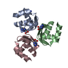
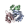
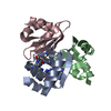
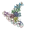
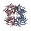
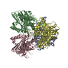
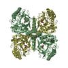
| Title | The Structure of the Intermediate Complex of the Arginine Repressor from Mycobacterium tuberculosis Bound to its DNA Operator and L-arginine. | ||||||
 Components Components |
| ||||||
 Keywords Keywords | TRANSCRIPTION REGULATOR / Mycobacterium tuberculosis / arginine repressor / DNA binding / ArgR-DNA complex / Structural Genomics / Mycobacterium Tuberculosis Structural Proteomics Project / XMTB / Amino-acid biosynthesis / Arginine biosynthesis / Cytoplasm / DNA-binding / Repressor / Transcription | ||||||
| Function / homology |  Function and homology information Function and homology informationregulation of arginine biosynthetic process / : / L-arginine biosynthetic process / arginine binding / protein complex oligomerization / cis-regulatory region sequence-specific DNA binding / transcription regulator complex / DNA-binding transcription factor activity / DNA binding / cytoplasm Similarity search - Function | ||||||
| Biological species |   Mycobacterium tuberculosis (bacteria) Mycobacterium tuberculosis (bacteria) | ||||||
| Method |  X-RAY DIFFRACTION / SYNCHROTRON / MOLECULAR REPLACEMENT / Resolution: 2.306 Å X-RAY DIFFRACTION / SYNCHROTRON / MOLECULAR REPLACEMENT / Resolution: 2.306 Å | ||||||
 Authors Authors | Cherney, L.T. / Cherney, M.M. / Garen, C.R. / James, M.N.G. / Mycobacterium Tuberculosis Structural Proteomics Project (XMTB) | ||||||
 Citation Citation | Journal: J.Mol.Biol. / Year: 2010 Title: crystal structure of the intermediate complex of the arginine repressor from Mycobacterium tuberculosis bound with its DNA operator reveals detailed mechanism of arginine repression. Authors: Cherney, L.T. / Cherney, M.M. / Garen, C.R. / James, M.N. | ||||||
| History |
|
- Structure visualization
| Structure viewer | Molecule: MolmilJmol/JSmol |
|---|
- Downloads & links
-Download
| PDBx/mmCIF format | 3laj.cif.gz | 240.9 KB | Display | PDBx/mmCIF format |
|---|---|---|---|---|
| PDB format | pdb3laj.ent.gz | 187.3 KB | Display | PDB format |
| PDBx/mmJSON format | 3laj.json.gz | Tree view | PDBx/mmJSON format | |
| Others |  Other downloads Other downloads |
-Validation report
| Arichive directory | https://data.pdbj.org/pub/pdb/validation_reports/la/3lajftp://data.pdbj.org/pub/pdb/validation_reports/la/3laj | HTTPS FTP |
|---|
-Related structure data
| Related structure data | 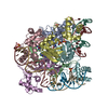 3lapC 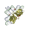 3ereS S: Starting model for refinement C: citing same article ( |
|---|---|
| Similar structure data |


-Links
 PDBj
PDBj


- Assembly
| Deposited unit | 
| ||||||||||||||||||||||||||||||||||||||||||||||||||||||||||||||||||||||||||||||||||||||||||||||||||||||
|---|---|---|---|---|---|---|---|---|---|---|---|---|---|---|---|---|---|---|---|---|---|---|---|---|---|---|---|---|---|---|---|---|---|---|---|---|---|---|---|---|---|---|---|---|---|---|---|---|---|---|---|---|---|---|---|---|---|---|---|---|---|---|---|---|---|---|---|---|---|---|---|---|---|---|---|---|---|---|---|---|---|---|---|---|---|---|---|---|---|---|---|---|---|---|---|---|---|---|---|---|---|---|---|
| 1 |
| ||||||||||||||||||||||||||||||||||||||||||||||||||||||||||||||||||||||||||||||||||||||||||||||||||||||
| Unit cell |
| ||||||||||||||||||||||||||||||||||||||||||||||||||||||||||||||||||||||||||||||||||||||||||||||||||||||
| Noncrystallographic symmetry (NCS) | NCS domain:
NCS domain segments:
NCS ensembles :
|
-Components
| #1: Protein | Mass: 17366.635 Da / Num. of mol.: 6 Source method: isolated from a genetically manipulated source Source: (gene. exp.) Mycobacterium tuberculosis (bacteria) / Strain: H37RV / Gene: ahrC, argR, MT1695, MTCY06H11.22, Rv1657 / Plasmid: PGST-1657 / Production host: #2: DNA chain | Mass: 4906.217 Da / Num. of mol.: 3 / Fragment: ARG box DNA segment, strand G / Source method: obtained synthetically #3: DNA chain | Mass: 4888.189 Da / Num. of mol.: 3 / Fragment: ARG box DNA segment, strand H / Source method: obtained synthetically #4: Chemical | ChemComp-ARG /   Type: L-peptide linking / Mass: 175.209 Da / Num. of mol.: 6 / Source method: obtained synthetically / Formula: C6H15N4O2 Type: L-peptide linking / Mass: 175.209 Da / Num. of mol.: 6 / Source method: obtained synthetically / Formula: C6H15N4O2#5: Water | ChemComp-HOH / |  Mass: 18.015 Da / Num. of mol.: 435 / Source method: isolated from a natural source / Formula: H2O Mass: 18.015 Da / Num. of mol.: 435 / Source method: isolated from a natural source / Formula: H2O |
|---|
-Experimental details
-Experiment
| Experiment | Method: X-RAY DIFFRACTION / Number of used crystals: 1 |
|---|
- Sample preparation
| Crystal | Density Matthews: 3.73 Å3/Da / Density % sol: 67.01 % |
|---|---|
| Crystal grow | Temperature: 295 K / Method: vapor diffusion / pH: 5.5 Details: 1M ammonium sulfate, 100 mM bis-tris buffer, 1% PEG 3350, pH 5.5, VAPOR DIFFUSION, temperature 295K |
-Data collection
| Diffraction | Mean temperature: 100 K |
|---|---|
| Diffraction source | Source: SYNCHROTRON / Site: ALS  / Beamline: 8.3.1 / Wavelength: 1.11587 Å / Beamline: 8.3.1 / Wavelength: 1.11587 Å |
| Detector | Type: ADSC QUANTUM 315 / Detector: CCD / Date: Feb 3, 2009 |
| Radiation | Protocol: SINGLE WAVELENGTH / Monochromatic (M) / Laue (L): M / Scattering type: x-ray |
| Radiation wavelength | Wavelength: 1.11587 Å / Relative weight: 1 |
| Reflection | Resolution: 2.3→50 Å / Num. all: 86698 / Num. obs: 84444 / % possible obs: 98.6 % / Observed criterion σ(F): 0 / Observed criterion σ(I): 0 / Redundancy: 2.2 % / Biso Wilson estimate: 58.3 Å2 / Rmerge(I) obs: 0.041 / Rsym value: 0.041 / Net I/σ(I): 16.6 |
| Reflection shell | Resolution: 2.3→2.38 Å / Redundancy: 2.1 % / Rmerge(I) obs: 0.85 / Mean I/σ(I) obs: 1.9 / Num. unique all: 8608 / Rsym value: 0.424 / % possible all: 98.6 |
- Processing
| Software |
| |||||||||||||||||||||||||||||||||||||||||||||||||||||||||||||||||||||||||||||||||||||||||||||||||||||||||||||||||||||||||||||||||||||||||||||||||||||||||||||||||||||||||||||||||||||||||||||||||||||||||||||||||||||||||
|---|---|---|---|---|---|---|---|---|---|---|---|---|---|---|---|---|---|---|---|---|---|---|---|---|---|---|---|---|---|---|---|---|---|---|---|---|---|---|---|---|---|---|---|---|---|---|---|---|---|---|---|---|---|---|---|---|---|---|---|---|---|---|---|---|---|---|---|---|---|---|---|---|---|---|---|---|---|---|---|---|---|---|---|---|---|---|---|---|---|---|---|---|---|---|---|---|---|---|---|---|---|---|---|---|---|---|---|---|---|---|---|---|---|---|---|---|---|---|---|---|---|---|---|---|---|---|---|---|---|---|---|---|---|---|---|---|---|---|---|---|---|---|---|---|---|---|---|---|---|---|---|---|---|---|---|---|---|---|---|---|---|---|---|---|---|---|---|---|---|---|---|---|---|---|---|---|---|---|---|---|---|---|---|---|---|---|---|---|---|---|---|---|---|---|---|---|---|---|---|---|---|---|---|---|---|---|---|---|---|---|---|---|---|---|---|---|---|---|
| Refinement | Method to determine structure: MOLECULAR REPLACEMENT Starting model: 3ere Resolution: 2.306→39.237 Å / SU ML: 0.27 / σ(F): 1.34 / σ(I): 0 / Stereochemistry target values: ML
| |||||||||||||||||||||||||||||||||||||||||||||||||||||||||||||||||||||||||||||||||||||||||||||||||||||||||||||||||||||||||||||||||||||||||||||||||||||||||||||||||||||||||||||||||||||||||||||||||||||||||||||||||||||||||
| Solvent computation | Shrinkage radii: 0.9 Å / VDW probe radii: 1.11 Å / Solvent model: FLAT BULK SOLVENT MODEL / Bsol: 66.496 Å2 / ksol: 0.359 e/Å3 | |||||||||||||||||||||||||||||||||||||||||||||||||||||||||||||||||||||||||||||||||||||||||||||||||||||||||||||||||||||||||||||||||||||||||||||||||||||||||||||||||||||||||||||||||||||||||||||||||||||||||||||||||||||||||
| Refinement step | Cycle: LAST / Resolution: 2.306→39.237 Å
| |||||||||||||||||||||||||||||||||||||||||||||||||||||||||||||||||||||||||||||||||||||||||||||||||||||||||||||||||||||||||||||||||||||||||||||||||||||||||||||||||||||||||||||||||||||||||||||||||||||||||||||||||||||||||
| Refine LS restraints |
| |||||||||||||||||||||||||||||||||||||||||||||||||||||||||||||||||||||||||||||||||||||||||||||||||||||||||||||||||||||||||||||||||||||||||||||||||||||||||||||||||||||||||||||||||||||||||||||||||||||||||||||||||||||||||
| Refine LS restraints NCS |
| |||||||||||||||||||||||||||||||||||||||||||||||||||||||||||||||||||||||||||||||||||||||||||||||||||||||||||||||||||||||||||||||||||||||||||||||||||||||||||||||||||||||||||||||||||||||||||||||||||||||||||||||||||||||||
| LS refinement shell |
|
