 Movie
Movie Controller

[English] 日本語
 Yorodumi
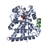
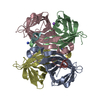
- PDB-3bue: Crystal structure of the C-terminal domain hexamer of ArgR from M... -
+ Open data

- Basic information
| Entry | Database: PDB / ID: 3bue | ||||||
|---|---|---|---|---|---|---|---|
| Title | Crystal structure of the C-terminal domain hexamer of ArgR from Mycobacterium tuberculosis | ||||||
 Components Components | Arginine repressor ArgR | ||||||
 Keywords Keywords | DNA BINDING PROTEIN / L-Arginine repressor protein / oligomerization domain / hexamer / L-arginine binding domain / Structural Genomics / TB Structural Genomics Consortium / TBSGC / Amino-acid biosynthesis / Arginine biosynthesis / DNA-binding / Repressor / Transcription / Transcription regulation / PSI-2 / Protein Structure Initiative | ||||||
| Function / homology |  Function and homology information Function and homology informationregulation of arginine biosynthetic process / : / L-arginine biosynthetic process / arginine binding / protein complex oligomerization / cis-regulatory region sequence-specific DNA binding / transcription regulator complex / DNA-binding transcription factor activity / DNA binding / cytoplasm Similarity search - Function | ||||||
| Biological species |   Mycobacterium tuberculosis (bacteria) Mycobacterium tuberculosis (bacteria) | ||||||
| Method |  X-RAY DIFFRACTION / SYNCHROTRON / MOLECULAR REPLACEMENT / Resolution: 2.15 Å X-RAY DIFFRACTION / SYNCHROTRON / MOLECULAR REPLACEMENT / Resolution: 2.15 Å | ||||||
 Authors Authors | Cherney, L.T. / Cherney, M.M. / Garen, C.R. / Lu, G.J. / James, M.N.G. / TB Structural Genomics Consortium (TBSGC) | ||||||
 Citation Citation | Journal: Acta Crystallogr.,Sect.D / Year: 2008 Title: Structure of the C-terminal domain of the arginine repressor protein from Mycobacterium tuberculosis. Authors: Cherney, L.T. / Cherney, M.M. / Garen, C.R. / Lu, G.J. / James, M.N. #1: Journal: Acta Crystallogr.,Sect.F / Year: 2007 Title: Expression, purification and preliminary X-ray analysis of the C-terminal domain of an arginine repressor protein from Mycobacterium tuberculosis. Authors: Lu, G.J. / Garen, C.R. / Cherney, M.M. / Cherney, L.T. / Lee, C. / James, M.N.G. | ||||||
| History |
|
- Structure visualization
| Structure viewer | Molecule: MolmilJmol/JSmol |
|---|
- Downloads & links
-Download
| PDBx/mmCIF format | 3bue.cif.gz | 100.7 KB | Display | PDBx/mmCIF format |
|---|---|---|---|---|
| PDB format | pdb3bue.ent.gz | 80 KB | Display | PDB format |
| PDBx/mmJSON format | 3bue.json.gz | Tree view | PDBx/mmJSON format | |
| Others |  Other downloads Other downloads |
-Validation report
| Arichive directory | https://data.pdbj.org/pub/pdb/validation_reports/bu/3bueftp://data.pdbj.org/pub/pdb/validation_reports/bu/3bue | HTTPS FTP |
|---|
-Related structure data
| Related structure data | 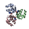 2zfzC 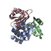 3cagC 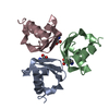 1b4bS S: Starting model for refinement C: citing same article ( |
|---|---|
| Similar structure data | |
| Other databases |


-Links
 PDBj
PDBj- Assembly
| Deposited unit | 
| ||||||||
|---|---|---|---|---|---|---|---|---|---|
| 1 |
| ||||||||
| 2 | 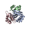
| ||||||||
| 3 | 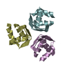
| ||||||||
| Unit cell |
| ||||||||
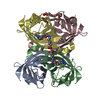
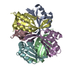
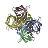
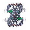
| Details | AUTHORS STATE THAT THE ASYMMETRIC UNIT CONTAINS ONE HEXAMER THAT IS A DIMER OF TRIMERS. EITHER HEXAMER OR TRIMER COULD BE THE BIOLOGICAL UNIT. |
-Components
| #1: Protein | Mass: 8137.206 Da / Num. of mol.: 6 / Fragment: C-terminal domain: Residues 92-170 Source method: isolated from a genetically manipulated source Source: (gene. exp.) Mycobacterium tuberculosis (bacteria) / Strain: H37Rv / Gene: argR, ahrC, Rv1657, MT1695, MTCY06H11.22 / Plasmid: pGST-1657 / Production host: #2: Water | ChemComp-HOH / |  Mass: 18.015 Da / Num. of mol.: 361 / Source method: isolated from a natural source / Formula: H2O Mass: 18.015 Da / Num. of mol.: 361 / Source method: isolated from a natural source / Formula: H2O |
|---|
-Experimental details
-Experiment
| Experiment | Method: X-RAY DIFFRACTION / Number of used crystals: 1 |
|---|
- Sample preparation
| Crystal | Density Matthews: 2.89 Å3/Da / Density % sol: 57.44 % |
|---|---|
| Crystal grow | Temperature: 293 K / Method: vapor diffusion, hanging drop / pH: 7.5 Details: Drops containing 1 microliter protein solution (10 mg/mL) and 0.5 microliter reservoir solution equilibrated against the reservoir solution (20% PEG 10000, 0.1 M HEPES pH 7.5), VAPOR ...Details: Drops containing 1 microliter protein solution (10 mg/mL) and 0.5 microliter reservoir solution equilibrated against the reservoir solution (20% PEG 10000, 0.1 M HEPES pH 7.5), VAPOR DIFFUSION, HANGING DROP, temperature 293K |
-Data collection
| Diffraction | Mean temperature: 100 K |
|---|---|
| Diffraction source | Source: SYNCHROTRON / Site: SSRL  / Beamline: BL7-1 / Wavelength: 0.97848 Å / Beamline: BL7-1 / Wavelength: 0.97848 Å |
| Detector | Type: ADSC QUANTUM 315 / Detector: CCD / Date: Mar 9, 2007 |
| Radiation | Protocol: SINGLE WAVELENGTH / Monochromatic (M) / Laue (L): M / Scattering type: x-ray |
| Radiation wavelength | Wavelength: 0.97848 Å / Relative weight: 1 |
| Reflection | Resolution: 2.15→50 Å / Num. all: 28214 / Num. obs: 28214 / % possible obs: 95 % / Observed criterion σ(F): 0 / Observed criterion σ(I): 0 / Redundancy: 1.8 % / Rsym value: 0.061 / Net I/σ(I): 11.77 |
| Reflection shell | Resolution: 2.15→2.23 Å / Redundancy: 1.7 % / Mean I/σ(I) obs: 2.26 / Rsym value: 0.252 / % possible all: 84 |
- Processing
| Software |
| ||||||||||||||||||||||||||||||||||||||||||||||||||||||||||||||||||||||||||||||||||||||||||||||||||||||||||||||||||||||||||||||||||||||||||||||||||||||||||||||||||||||||||
|---|---|---|---|---|---|---|---|---|---|---|---|---|---|---|---|---|---|---|---|---|---|---|---|---|---|---|---|---|---|---|---|---|---|---|---|---|---|---|---|---|---|---|---|---|---|---|---|---|---|---|---|---|---|---|---|---|---|---|---|---|---|---|---|---|---|---|---|---|---|---|---|---|---|---|---|---|---|---|---|---|---|---|---|---|---|---|---|---|---|---|---|---|---|---|---|---|---|---|---|---|---|---|---|---|---|---|---|---|---|---|---|---|---|---|---|---|---|---|---|---|---|---|---|---|---|---|---|---|---|---|---|---|---|---|---|---|---|---|---|---|---|---|---|---|---|---|---|---|---|---|---|---|---|---|---|---|---|---|---|---|---|---|---|---|---|---|---|---|---|---|---|
| Refinement | Method to determine structure: MOLECULAR REPLACEMENT Starting model: PDB entry 1B4B Resolution: 2.15→44.81 Å / Cor.coef. Fo:Fc: 0.957 / Cor.coef. Fo:Fc free: 0.911 / SU B: 4.988 / SU ML: 0.128 / Cross valid method: THROUGHOUT / ESU R: 0.212 / ESU R Free: 0.196 / Stereochemistry target values: MAXIMUM LIKELIHOOD / Details: HYDROGENS HAVE BEEN ADDED IN THE RIDING POSITIONS
| ||||||||||||||||||||||||||||||||||||||||||||||||||||||||||||||||||||||||||||||||||||||||||||||||||||||||||||||||||||||||||||||||||||||||||||||||||||||||||||||||||||||||||
| Solvent computation | Ion probe radii: 0.8 Å / Shrinkage radii: 0.8 Å / VDW probe radii: 1.2 Å / Solvent model: MASK | ||||||||||||||||||||||||||||||||||||||||||||||||||||||||||||||||||||||||||||||||||||||||||||||||||||||||||||||||||||||||||||||||||||||||||||||||||||||||||||||||||||||||||
| Displacement parameters | Biso mean: 25.627 Å2
| ||||||||||||||||||||||||||||||||||||||||||||||||||||||||||||||||||||||||||||||||||||||||||||||||||||||||||||||||||||||||||||||||||||||||||||||||||||||||||||||||||||||||||
| Refinement step | Cycle: LAST / Resolution: 2.15→44.81 Å
| ||||||||||||||||||||||||||||||||||||||||||||||||||||||||||||||||||||||||||||||||||||||||||||||||||||||||||||||||||||||||||||||||||||||||||||||||||||||||||||||||||||||||||
| Refine LS restraints |
| ||||||||||||||||||||||||||||||||||||||||||||||||||||||||||||||||||||||||||||||||||||||||||||||||||||||||||||||||||||||||||||||||||||||||||||||||||||||||||||||||||||||||||
| LS refinement shell | Resolution: 2.15→2.21 Å / Total num. of bins used: 20
|
