THE FAE ATOM OF T13 IS MISSING IN RESIDUE B1241 AND D1241 CAUSED BY COVALENT BINDING OF T13 TO CYS 241
Sequence details
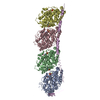
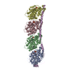
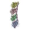
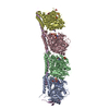
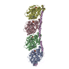
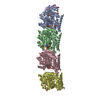
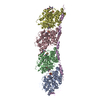
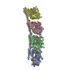
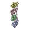
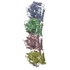
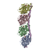
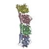
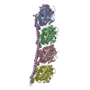
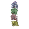
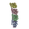
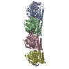
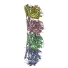
THERE IS ONE COMPLEX IN THE ASYMMETRIC UNIT, WHICH CONSISTS OF TWO ALPHA-BETA TUBULIN HETERODIMERS ...THERE IS ONE COMPLEX IN THE ASYMMETRIC UNIT, WHICH CONSISTS OF TWO ALPHA-BETA TUBULIN HETERODIMERS (CHAINS A-B AND C-D), AND ONE STATHMIN-LIKE DOMAIN OF RB3 (RB3-SLD) WHICH CORRESPONDS TO STAHMIN RESIDUES 5 TO 145 WITH THE ADDITION OF ONE ACETYLATED ALANINE AT THE N-TERMINUS. THE NUMBERING OF RB3-SLD IS ACCORDING TO THE STATHMIN SEQUENCE. ALPHA-TUBULIN AND BETA-TUBULIN HAVE BEEN ALIGNED AS IN NOGALES ET AL., NATURE VOL 391,199-203. IN THIS ALIGNMENT, RESIDUES 45-46 AND 361-368 OF ALPHA-TUBULIN ARE MISSING IN BETA-TUBULIN. AS THE SEQUENCE OF OVIS ARIES(SHEEP) TUBULIN IS NOT AVAILABLE, THE BOS TAURUS TUBULIN SEQUENCES (ALPHA: ISOTYPE 1A, GI:194666935, BETA: ISOTYPE 2, GI:51491829) WERE USED AS A REFERENCE BUT FOR THE ILE TO VAL SUBSTITUTION AT POSITION 318 ON BETA TUBULIN. THIS IS BASED ON DIFFERENCES BETWEEN TUBULIN ISOTYPES AND ON THE RELATIVE EXPRESSION ON THESE ISOTYPES IN MAMMALIAN BRAIN.
-
Experimental details
-
Experiment
Experiment
Method: X-RAY DIFFRACTION / Number of used crystals: 1
-
Sample preparation
Crystal
Density Matthews: 3.84 Å3/Da / Density % sol: 68 %
Crystal grow
Temperature: 277 K / Method: vapor diffusion, hanging drop / pH: 7 Details: PEG, PIPES BUFFER, pH 7.00, VAPOR DIFFUSION, HANGING DROP, temperature 277K
In the structure databanks used in Yorodumi, some data are registered as the other names, "COVID-19 virus" and "2019-nCoV". Here are the details of the virus and the list of structure data.
Jan 31, 2019. EMDB accession codes are about to change! (news from PDBe EMDB page)
EMDB accession codes are about to change! (news from PDBe EMDB page)
The allocation of 4 digits for EMDB accession codes will soon come to an end. Whilst these codes will remain in use, new EMDB accession codes will include an additional digit and will expand incrementally as the available range of codes is exhausted. The current 4-digit format prefixed with “EMD-” (i.e. EMD-XXXX) will advance to a 5-digit format (i.e. EMD-XXXXX), and so on. It is currently estimated that the 4-digit codes will be depleted around Spring 2019, at which point the 5-digit format will come into force.
The EM Navigator/Yorodumi systems omit the EMD- prefix.
Related info.:Q: What is EMD? / ID/Accession-code notation in Yorodumi/EM Navigator
Yorodumi is a browser for structure data from EMDB, PDB, SASBDB, etc.
This page is also the successor to EM Navigator detail page, and also detail information page/front-end page for Omokage search.
The word "yorodu" (or yorozu) is an old Japanese word meaning "ten thousand". "mi" (miru) is to see.
Related info.:EMDB / PDB / SASBDB / Comparison of 3 databanks / Yorodumi Search / Aug 31, 2016. New EM Navigator & Yorodumi / Yorodumi Papers / Jmol/JSmol / Function and homology information / Changes in new EM Navigator and Yorodumi
 Movie
Movie Controller


 Open data
 Basic information
 Components
 Keywords
 Function and homology information
 X-RAY DIFFRACTION /
  Authors
 Citation
 Structure visualization
 Downloads & links
 Other downloads


 PDBj
PDBj


 Assembly
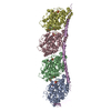


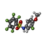

 Mass: 523.180 Da / Num. of mol.: 2 / Source method: obtained synthetically / Formula: C10H16N5O14P3 / Comment: GTP, energy-carrying molecule*YM
 Mass: 24.305 Da / Num. of mol.: 3 / Source method: obtained synthetically / Formula: Mg
 Mass: 371.255 Da / Num. of mol.: 4 / Source method: obtained synthetically / Formula: C13H7F6NO3S
 Type: RNA linking / Mass: 443.201 Da / Num. of mol.: 2 / Source method: obtained synthetically / Formula: C10H15N5O11P2 / Comment: GDP, energy-carrying molecule*YM
 Sample preparation
 / Beamline: ID14-4 / Wavelength: 0.978 / Wavelength: 0.978 Å
 Processing