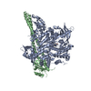
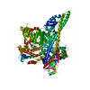
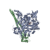
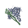
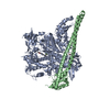
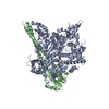
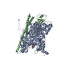
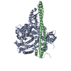
登録情報 データベース : PDB / ID : 3hizタイトル Crystal structure of p110alpha H1047R mutant in complex with niSH2 of p85alpha Phosphatidylinositol 3-kinase regulatory subunit alpha Phosphatidylinositol-4,5-bisphosphate 3-kinase catalytic subunit alpha isoform キーワード / / / / / / / / / / / / / / / / / / / / / / / 機能・相同性 分子機能 ドメイン・相同性 構成要素
/ / / / / / / / / / / / / / / / / / / / / / / / / / / / / / / / / / / / / / / / / / / / / / / / / / / / / / / / / / / / / / / / / / / / / / / / / / / / / / / / / / / / / / / / / / / / / / / / / / / / / / / / / / / / / / / / / / / / / / / / / / / / / / / / / / / / / / / / / / / / / / / / / / / / / / / / / / / / / / / / / / / / / / / / / / / / / / / / / / / / / / / / / / / / / / 生物種 Homo sapiens (ヒト)手法 / / / 解像度 : 3.3 Å データ登録者 Amzel, L.M. / Vogelstein, B. / Gabelli, S.B. / Mandelker, D. ジャーナル : Proc.Natl.Acad.Sci.USA / 年 : 2009タイトル : A frequent kinase domain mutation that changes the interaction between PI3K{alpha} and the membrane.著者 : Mandelker, D. / Gabelli, S.B. / Schmidt-Kittler, O. / Zhu, J. / Cheong, I. / Huang, C.H. / Kinzler, K.W. / Vogelstein, B. / Amzel, L.M. 履歴 登録 2009年5月20日 登録サイト / 処理サイト 改定 1.0 2009年9月29日 Provider / タイプ 改定 1.1 2011年7月13日 Group 改定 1.2 2017年11月1日 Group / カテゴリ 改定 1.3 2021年10月13日 Group / カテゴリ / struct_ref_seq_difItem / _database_2.pdbx_database_accession / _struct_ref_seq_dif.details改定 1.4 2023年9月6日 Group / Refinement descriptionカテゴリ / chem_comp_bond / pdbx_initial_refinement_model
すべて表示 表示を減らす
 ムービー
ムービー コントローラー


 データを開く
 基本情報
基本情報 要素
要素 キーワード
キーワード 機能・相同性情報
 Homo sapiens (ヒト)
 X線回折 /
  データ登録者
 引用
引用 構造の表示
構造の表示 ダウンロードとリンク
 その他のダウンロード


 PDBj
PDBj


 集合体
集合体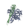

 Spodoptera frugiperda (ツマジロクサヨトウ)
 試料調製
試料調製 / ビームライン: 31-ID / 波長: 0.97929 Å
 解析
解析