Protocol: SINGLE WAVELENGTH / Scattering type: x-ray
Radiation wavelength
Wavelength: 0.9788 Å / Relative weight: 1
Reflection
Resolution: 3.36→50 Å / Num. obs: 28636 / % possible obs: 99.9 % / Redundancy: 9 % / Rmerge(I) obs: 0.103 / Χ2: 2.127 / Net I/av σ(I): 35.706 / Net I/σ(I): 10.9 / Num. measured all: 256465
Reflection shell
Diffraction-ID: 1 / Rejects: _
Resolution (Å)
Redundancy (%)
Rmerge(I) obs
Num. unique all
Χ2
% possible all
3.36-3.42
9.2
0.712
1400
1.748
100
3.42-3.48
9.2
0.644
1405
2.283
100
3.48-3.55
9.1
0.49
1424
1.68
100
3.55-3.62
9.2
0.407
1416
1.75
100
3.62-3.7
9.2
0.336
1396
1.802
100
3.7-3.78
9.2
0.28
1418
1.843
100
3.78-3.88
9.2
0.232
1418
1.886
100
3.88-3.98
9.1
0.195
1405
1.907
100
3.98-4.1
9.1
0.17
1413
1.941
100
4.1-4.23
9.1
0.141
1428
2.024
100
4.23-4.38
9.1
0.126
1425
2.201
100
4.38-4.56
9
0.103
1413
2.264
100
4.56-4.77
9
0.096
1431
2.407
100
4.77-5.02
8.9
0.097
1424
2.532
100
5.02-5.33
8.9
0.096
1435
2.651
100
5.33-5.74
8.8
0.101
1441
2.588
100
5.74-6.32
8.8
0.097
1452
2.42
100
6.32-7.23
8.6
0.076
1463
2.302
100
7.23-9.1
8.5
0.045
1478
2.185
99.9
9.1-50
8
0.036
1551
2.175
98.4
-
Processing
Software
Name
Version
Classification
NB
REFMAC
5.6.0117
refinement
PDB_EXTRACT
3.14
dataextraction
DENZO
datareduction
SCALEPACK
datascaling
Refinement
Resolution: 3.5→91.51 Å / Cor.coef. Fo:Fc: 0.918 / Cor.coef. Fo:Fc free: 0.821 / WRfactor Rfree: 0.3388 / WRfactor Rwork: 0.2367 / FOM work R set: 0.7462 / SU B: 90.419 / SU ML: 0.643 / SU R Cruickshank DPI: 0.5687 / SU Rfree: 0.8008 / Cross valid method: THROUGHOUT / σ(F): 0 / ESU R Free: 0.801 / Stereochemistry target values: MAXIMUM LIKELIHOOD Details: HYDROGENS HAVE BEEN USED IF PRESENT IN THE INPUT U VALUES : RESIDUAL ONLY
Rfactor
Num. reflection
% reflection
Selection details
Rfree
0.3388
1316
5.1 %
RANDOM
Rwork
0.2367
24266
-
-
obs
0.2418
25582
99.84 %
-
Solvent computation
Ion probe radii: 0.8 Å / Shrinkage radii: 0.8 Å / VDW probe radii: 1.4 Å / Solvent model: MASK
In the structure databanks used in Yorodumi, some data are registered as the other names, "COVID-19 virus" and "2019-nCoV". Here are the details of the virus and the list of structure data.
Jan 31, 2019. EMDB accession codes are about to change! (news from PDBe EMDB page)
EMDB accession codes are about to change! (news from PDBe EMDB page)
The allocation of 4 digits for EMDB accession codes will soon come to an end. Whilst these codes will remain in use, new EMDB accession codes will include an additional digit and will expand incrementally as the available range of codes is exhausted. The current 4-digit format prefixed with “EMD-” (i.e. EMD-XXXX) will advance to a 5-digit format (i.e. EMD-XXXXX), and so on. It is currently estimated that the 4-digit codes will be depleted around Spring 2019, at which point the 5-digit format will come into force.
The EM Navigator/Yorodumi systems omit the EMD- prefix.
Related info.:Q: What is EMD? / ID/Accession-code notation in Yorodumi/EM Navigator
Yorodumi is a browser for structure data from EMDB, PDB, SASBDB, etc.
This page is also the successor to EM Navigator detail page, and also detail information page/front-end page for Omokage search.
The word "yorodu" (or yorozu) is an old Japanese word meaning "ten thousand". "mi" (miru) is to see.
Related info.:EMDB / PDB / SASBDB / Comparison of 3 databanks / Yorodumi Search / Aug 31, 2016. New EM Navigator & Yorodumi / Yorodumi Papers / Jmol/JSmol / Function and homology information / Changes in new EM Navigator and Yorodumi
 Movie
Movie Controller


 Open data
 Basic information
 Components
 Keywords
 Function and homology information
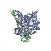
 Homo sapiens (human)
 X-RAY DIFFRACTION /
  Authors
 United States, 1items
  Citation
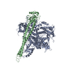
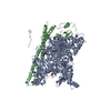
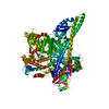
 Structure visualization
 Downloads & links
 Other downloads

 PDBj
PDBj


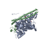
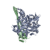
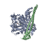
 Assembly

 Spodoptera frugiperda (fall armyworm)
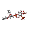
 Mass: 634.354 Da / Num. of mol.: 2 / Source method: obtained synthetically / Formula: C17H33O19P3
 Mass: 94.971 Da / Num. of mol.: 1 / Source method: obtained synthetically / Formula: PO4
 Mass: 18.015 Da / Num. of mol.: 4 / Source method: isolated from a natural source / Formula: H2O
 Sample preparation
 Processing