 Movie
Movie Controller

[English] 日本語
 Yorodumi
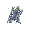
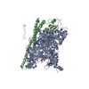
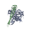
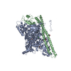
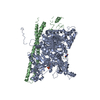
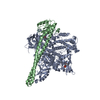
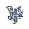
- PDB-3hhm: Crystal structure of p110alpha H1047R mutant in complex with niSH... -
+ Open data

- Basic information
| Entry | Database: PDB / ID: 3hhm | ||||||
|---|---|---|---|---|---|---|---|
| Title | Crystal structure of p110alpha H1047R mutant in complex with niSH2 of p85alpha and the drug wortmannin | ||||||
 Components Components |
| ||||||
 Keywords Keywords | TRANSFERASE/ONCOPROTEIN / p110 / p85 / PI3KCA / PI3K / PIK3R1 / phosphatidilynositol 3 / 4 / 5-triphosphate / wortmannin / H1047R / ATP-binding / Disease mutation / Kinase / Nucleotide-binding / Oncogene / Polymorphism / Transferase / TRANSFERASE-ONCOPROTEIN COMPLEX | ||||||
| Function / homology |  Function and homology information Function and homology informationperinuclear endoplasmic reticulum membrane / regulation of toll-like receptor 4 signaling pathway / response to muscle inactivity / phosphatidylinositol kinase activity / phosphatidylinositol 3-kinase regulator activity / 1-phosphatidylinositol-3-kinase regulator activity / positive regulation of endoplasmic reticulum unfolded protein response / regulation of actin filament organization / phosphatidylinositol 3-kinase activator activity / negative regulation of actin filament depolymerization ...perinuclear endoplasmic reticulum membrane / regulation of toll-like receptor 4 signaling pathway / response to muscle inactivity / phosphatidylinositol kinase activity / phosphatidylinositol 3-kinase regulator activity / 1-phosphatidylinositol-3-kinase regulator activity / positive regulation of endoplasmic reticulum unfolded protein response / regulation of actin filament organization / phosphatidylinositol 3-kinase activator activity / negative regulation of actin filament depolymerization / response to butyrate / T follicular helper cell differentiation / IRS-mediated signalling / interleukin-18-mediated signaling pathway / myeloid leukocyte migration / phosphatidylinositol 3-kinase regulatory subunit binding / response to L-leucine / PI3K events in ERBB4 signaling / neurotrophin TRKA receptor binding / positive regulation of focal adhesion disassembly / cellular response to hydrostatic pressure / autosome genomic imprinting / cis-Golgi network / Activated NTRK2 signals through PI3K / negative regulation of fibroblast apoptotic process / ErbB-3 class receptor binding / transmembrane receptor protein tyrosine kinase adaptor activity / negative regulation of stress fiber assembly / Activated NTRK3 signals through PI3K / phosphatidylinositol 3-kinase complex, class IB / phosphatidylinositol 3-kinase complex / Co-stimulation by ICOS / TORC2 signaling / RHOD GTPase cycle / positive regulation of protein localization to membrane / Signaling by cytosolic FGFR1 fusion mutants / vasculature development / regulation of cellular respiration / Nephrin family interactions / RHOF GTPase cycle / kinase activator activity / Signaling by LTK in cancer / 1-phosphatidylinositol-4-phosphate 3-kinase activity / Signaling by LTK / anoikis / RND1 GTPase cycle / RND2 GTPase cycle / positive regulation of leukocyte migration / RND3 GTPase cycle / relaxation of cardiac muscle / phosphatidylinositol 3-kinase complex, class IA / positive regulation of filopodium assembly / MET activates PI3K/AKT signaling / PI3K/AKT activation / phosphatidylinositol-4,5-bisphosphate 3-kinase / 1-phosphatidylinositol-4,5-bisphosphate 3-kinase activity / growth hormone receptor signaling pathway / phosphatidylinositol 3-kinase / insulin binding / phosphatidylinositol-3-phosphate biosynthetic process / Signaling by ALK / cardiac muscle cell contraction / RHOV GTPase cycle / 1-phosphatidylinositol-3-kinase activity / vascular endothelial growth factor signaling pathway / RHOB GTPase cycle / natural killer cell mediated cytotoxicity / GP1b-IX-V activation signalling / Erythropoietin activates Phosphoinositide-3-kinase (PI3K) / PI-3K cascade:FGFR3 / response to dexamethasone / PI-3K cascade:FGFR2 / PI-3K cascade:FGFR4 / negative regulation of macroautophagy / PI-3K cascade:FGFR1 / RHOJ GTPase cycle / RHOC GTPase cycle / negative regulation of osteoclast differentiation / phosphatidylinositol phosphate biosynthetic process / phosphatidylinositol-mediated signaling / Synthesis of PIPs at the plasma membrane / RHOU GTPase cycle / CDC42 GTPase cycle / RET signaling / negative regulation of anoikis / T cell differentiation / Interleukin-3, Interleukin-5 and GM-CSF signaling / PI3K events in ERBB2 signaling / RHOG GTPase cycle / insulin receptor substrate binding / negative regulation of cell-matrix adhesion / intercalated disc / PI3K Cascade / extrinsic apoptotic signaling pathway via death domain receptors / Role of LAT2/NTAL/LAB on calcium mobilization / regulation of multicellular organism growth / RAC3 GTPase cycle / RHOA GTPase cycle / CD28 dependent PI3K/Akt signaling / RAC2 GTPase cycle Similarity search - Function | ||||||
| Biological species |  Homo sapiens (human) Homo sapiens (human) | ||||||
| Method |  X-RAY DIFFRACTION / SYNCHROTRON / FOURIER SYNTHESIS / Resolution: 2.8 Å X-RAY DIFFRACTION / SYNCHROTRON / FOURIER SYNTHESIS / Resolution: 2.8 Å | ||||||
 Authors Authors | Amzel, L.M. / Vogelstein, B. / Gabelli, S.B. / Mandelker, D. | ||||||
 Citation Citation | Journal: Proc.Natl.Acad.Sci.USA / Year: 2009 Title: A frequent kinase domain mutation that changes the interaction between PI3K{alpha} and the membrane. Authors: Mandelker, D. / Gabelli, S.B. / Schmidt-Kittler, O. / Zhu, J. / Cheong, I. / Huang, C.H. / Kinzler, K.W. / Vogelstein, B. / Amzel, L.M. | ||||||
| History |
|
- Structure visualization
| Structure viewer | Molecule: MolmilJmol/JSmol |
|---|
- Downloads & links
-Download
| PDBx/mmCIF format | 3hhm.cif.gz | 278.8 KB | Display | PDBx/mmCIF format |
|---|---|---|---|---|
| PDB format | pdb3hhm.ent.gz | 217.6 KB | Display | PDB format |
| PDBx/mmJSON format | 3hhm.json.gz | Tree view | PDBx/mmJSON format | |
| Others |  Other downloads Other downloads |
-Validation report
| Arichive directory | https://data.pdbj.org/pub/pdb/validation_reports/hh/3hhmftp://data.pdbj.org/pub/pdb/validation_reports/hh/3hhm | HTTPS FTP |
|---|
-Related structure data
| Related structure data |  3hizC  2rd0S C: citing same article ( S: Starting model for refinement |
|---|---|
| Similar structure data |


-Links
 PDBj
PDBj


- Assembly
| Deposited unit | 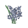
| ||||||||
|---|---|---|---|---|---|---|---|---|---|
| 1 |
| ||||||||
| Unit cell |
|
-Components
| #1: Protein | Mass: 127294.086 Da / Num. of mol.: 1 / Mutation: H1047R Source method: isolated from a genetically manipulated source Source: (gene. exp.) Homo sapiens (human) / Gene: PIK3CA / Plasmid: pFastbac HT-A / Production host:   Spodoptera frugiperda (fall armyworm) Spodoptera frugiperda (fall armyworm)References: UniProt: P42336, phosphatidylinositol-4,5-bisphosphate 3-kinase |
|---|---|
| #2: Protein | Mass: 44262.496 Da / Num. of mol.: 1 / Fragment: UNP residues 322-694 / Mutation: D330N Source method: isolated from a genetically manipulated source Source: (gene. exp.) Homo sapiens (human) / Gene: PIK3R1 / Plasmid: pFastbac HT-A / Production host: Spodoptera frugiperda (fall armyworm) / References: UniProt: P27986 |
| #3: Chemical | ChemComp-KWT / (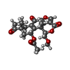  Mass: 428.432 Da / Num. of mol.: 1 / Source method: obtained synthetically / Formula: C23H24O8 Mass: 428.432 Da / Num. of mol.: 1 / Source method: obtained synthetically / Formula: C23H24O8 |
| #4: Water | ChemComp-HOH /  Mass: 18.015 Da / Num. of mol.: 115 / Source method: isolated from a natural source / Formula: H2O Mass: 18.015 Da / Num. of mol.: 115 / Source method: isolated from a natural source / Formula: H2O |
-Experimental details
-Experiment
| Experiment | Method: X-RAY DIFFRACTION / Number of used crystals: 1 |
|---|
- Sample preparation
| Crystal | Density Matthews: 3.11 Å3/Da / Density % sol: 60.48 % |
|---|---|
| Crystal grow | Temperature: 298 K / Method: vapor diffusion, hanging drop / pH: 6.8 Details: Na formate, pH 6.8, VAPOR DIFFUSION, HANGING DROP, temperature 298K |
-Data collection
| Diffraction | Mean temperature: 100 K | ||||||||||||||||||||||||||||||||||||||||||||||||||||||||||||||||||
|---|---|---|---|---|---|---|---|---|---|---|---|---|---|---|---|---|---|---|---|---|---|---|---|---|---|---|---|---|---|---|---|---|---|---|---|---|---|---|---|---|---|---|---|---|---|---|---|---|---|---|---|---|---|---|---|---|---|---|---|---|---|---|---|---|---|---|---|
| Diffraction source | Source: SYNCHROTRON / Site: APS  / Beamline: 31-ID / Wavelength: 0.97929 Å / Beamline: 31-ID / Wavelength: 0.97929 Å | ||||||||||||||||||||||||||||||||||||||||||||||||||||||||||||||||||
| Detector | Type: MAR CCD 165 mm / Detector: CCD / Date: Mar 3, 2009 | ||||||||||||||||||||||||||||||||||||||||||||||||||||||||||||||||||
| Radiation | Protocol: SINGLE WAVELENGTH / Monochromatic (M) / Laue (L): M / Scattering type: x-ray | ||||||||||||||||||||||||||||||||||||||||||||||||||||||||||||||||||
| Radiation wavelength | Wavelength: 0.97929 Å / Relative weight: 1 | ||||||||||||||||||||||||||||||||||||||||||||||||||||||||||||||||||
| Reflection | Resolution: 2.8→50 Å / Num. obs: 53371 / % possible obs: 99.8 % / Redundancy: 6.8 % / Rmerge(I) obs: 0.093 / Χ2: 1.105 / Net I/σ(I): 23.607 | ||||||||||||||||||||||||||||||||||||||||||||||||||||||||||||||||||
| Reflection shell |
|
- Processing
| Software |
| ||||||||||||||||||||||||||||
|---|---|---|---|---|---|---|---|---|---|---|---|---|---|---|---|---|---|---|---|---|---|---|---|---|---|---|---|---|---|
| Refinement | Method to determine structure: FOURIER SYNTHESIS Starting model: PDB entry 2RD0 Resolution: 2.8→50 Å / WRfactor Rfree: 0.283 / WRfactor Rwork: 0.223 / Occupancy max: 1 / Occupancy min: 1 / FOM work R set: 0.75 / SU R Cruickshank DPI: 0.812 / SU Rfree: 0.404 / Isotropic thermal model: ISOTROPIC / Cross valid method: THROUGHOUT / σ(I): 0 / Stereochemistry target values: Engh & Huber
| ||||||||||||||||||||||||||||
| Displacement parameters | Biso max: 227.75 Å2 / Biso mean: 66.68 Å2 / Biso min: 26.12 Å2 | ||||||||||||||||||||||||||||
| Refinement step | Cycle: LAST / Resolution: 2.8→50 Å
|
