 Movie
Movie Controller

[English] 日本語
 Yorodumi
- PDB-3h4g: Structure of aldehyde reductase holoenzyme in complex with potent... -
+ Open data

- Basic information
| Entry | Database: PDB / ID: 3h4g | |||||||||
|---|---|---|---|---|---|---|---|---|---|---|
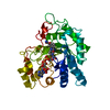
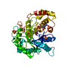
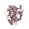
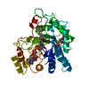
| Title | Structure of aldehyde reductase holoenzyme in complex with potent aldose reductase inhibitor Fidarestat: Implications for inhibitor binding and selectivity | |||||||||
 Components Components | Alcohol dehydrogenase [NADP+] | |||||||||
 Keywords Keywords |  OXIDOREDUCTASE / TIM BARREL / ALDO-KETO REDUCTASE / TERNARY COMPLEX / NADP OXIDOREDUCTASE / TIM BARREL / ALDO-KETO REDUCTASE / TERNARY COMPLEX / NADP | |||||||||
| Function / homology |  Function and homology informationglucuronate reductase / glucuronolactone reductase / glucuronolactone reductase activity / methylglyoxal reductase (NADPH) (acetol producing) activity / D-glucuronate catabolic process / alcohol dehydrogenase (NADP+) / aldehyde catabolic process / cellular detoxification of aldehyde / L-glucuronate reductase activity / glycerol dehydrogenase [NADP+] activity ...glucuronate reductase / glucuronolactone reductase / glucuronolactone reductase activity / methylglyoxal reductase (NADPH) (acetol producing) activity / D-glucuronate catabolic process / alcohol dehydrogenase (NADP+) / aldehyde catabolic process / cellular detoxification of aldehyde / L-glucuronate reductase activity / glycerol dehydrogenase [NADP+] activity / D/L-glyceraldehyde reductase / L-ascorbic acid biosynthetic process / allyl-alcohol dehydrogenase / allyl-alcohol dehydrogenase activity / aldose reductase (NADPH) activity / lipid metabolic process / apical plasma membrane / cytosol Function and homology informationglucuronate reductase / glucuronolactone reductase / glucuronolactone reductase activity / methylglyoxal reductase (NADPH) (acetol producing) activity / D-glucuronate catabolic process / alcohol dehydrogenase (NADP+) / aldehyde catabolic process / cellular detoxification of aldehyde / L-glucuronate reductase activity / glycerol dehydrogenase [NADP+] activity ...glucuronate reductase / glucuronolactone reductase / glucuronolactone reductase activity / methylglyoxal reductase (NADPH) (acetol producing) activity / D-glucuronate catabolic process / alcohol dehydrogenase (NADP+) / aldehyde catabolic process / cellular detoxification of aldehyde / L-glucuronate reductase activity / glycerol dehydrogenase [NADP+] activity / D/L-glyceraldehyde reductase / L-ascorbic acid biosynthetic process / allyl-alcohol dehydrogenase / allyl-alcohol dehydrogenase activity / aldose reductase (NADPH) activity / lipid metabolic process / apical plasma membrane / cytosolSimilarity search - Function | |||||||||
| Biological species |  Sus scrofa (pig) Sus scrofa (pig) | |||||||||
| Method | X-RAY DIFFRACTION / SYNCHROTRON / MOLECULAR REPLACEMENT / Resolution: 1.85 Å | |||||||||
 Authors Authors | Carbone, V. / El-Kabbani, O. | |||||||||
 Citation Citation | Journal: J.Med.Chem. / Year: 2005 Title: Structure of aldehyde reductase holoenzyme in complex with the potent aldose reductase inhibitor fidarestat: implications for inhibitor binding and selectivity Authors: El-Kabbani, O. / Carbone, V. / Darmanin, C. / Oka, M. / Mitschler, A. / Podjarny, A. / Schulze-Briese, C. / Chung, R.P. | |||||||||
| History |
|
- Structure visualization
| Structure viewer | Molecule: MolmilJmol/JSmol |
|---|
- Downloads & links
-Download
| PDBx/mmCIF format | 3h4g.cif.gz | 88.5 KB | Display | PDBx/mmCIF format |
|---|---|---|---|---|
| PDB format | pdb3h4g.ent.gz | 64.8 KB | Display | PDB format |
| PDBx/mmJSON format | 3h4g.json.gz | Tree view | PDBx/mmJSON format | |
| Others |  Other downloads Other downloads |
-Validation report
| Arichive directory | https://data.pdbj.org/pub/pdb/validation_reports/h4/3h4gftp://data.pdbj.org/pub/pdb/validation_reports/h4/3h4g | HTTPS FTP |
|---|
-Related structure data
| Related structure data |  2a0o S: Starting model for refinement |
|---|---|
| Similar structure data |


-Links
 PDBj
PDBj
- Assembly
| Deposited unit | 
| ||||||||||||
|---|---|---|---|---|---|---|---|---|---|---|---|---|---|
| 1 |
| ||||||||||||
| Unit cell |
| ||||||||||||
| Components on special symmetry positions |
|
-Components
| #1: Protein | Mass: 36626.953 Da / Num. of mol.: 1 / Source method: isolated from a natural source / Source: (natural) Sus scrofa (pig) / Tissue: kidney / References: UniProt: P50578, alcohol dehydrogenase (NADP+) |
|---|---|
| #2: Chemical | ChemComp-SO4 / Sulfate  Mass: 96.063 Da / Num. of mol.: 1 / Source method: obtained synthetically / Formula: SO4 Mass: 96.063 Da / Num. of mol.: 1 / Source method: obtained synthetically / Formula: SO4 |
| #3: Chemical | ChemComp-NAP / Nicotinamide adenine dinucleotide phosphate  Mass: 743.405 Da / Num. of mol.: 1 / Source method: obtained synthetically / Formula: C21H28N7O17P3 Mass: 743.405 Da / Num. of mol.: 1 / Source method: obtained synthetically / Formula: C21H28N7O17P3 |
| #4: Chemical | ChemComp-FID / (Fidarestat  Mass: 279.224 Da / Num. of mol.: 1 / Source method: obtained synthetically / Formula: C12H10FN3O4 / Comment: inhibitor*YM Mass: 279.224 Da / Num. of mol.: 1 / Source method: obtained synthetically / Formula: C12H10FN3O4 / Comment: inhibitor*YM |
| #5: Water | ChemComp-HOH / Water Mass: 18.015 Da / Num. of mol.: 255 / Source method: isolated from a natural source / Formula: H2O Mass: 18.015 Da / Num. of mol.: 255 / Source method: isolated from a natural source / Formula: H2O |
| Sequence details | THE PUBLISHED SEQUENCE IS FROM EXPRESSED MATERIAL WHILE THE SEQUENCE FOR THIS ENTRY IS FROM ...THE PUBLISHED SEQUENCE IS FROM EXPRESSED MATERIAL WHILE THE SEQUENCE FOR THIS ENTRY IS FROM PURIFIED MATERIAL. O.EL-KABBANI,N.C.GREEN,G.LIN,M.CARSON,S.V.L.NARAYANA, K.M.MOORE,T.G.FLYNN,L.J.DELUCAS. STRUCTURES |
-Experimental details
-Experiment
| Experiment | Method: X-RAY DIFFRACTION / Number of used crystals: 1 |
|---|
- Sample preparation
| Crystal | Density Matthews: 2.18 Å3/Da / Density % sol: 43.69 % |
|---|---|
| Crystal grow | Temperature: 295 K / Method: vapor diffusion, hanging drop / pH: 8.1 Details: 2M ammonium sulphate, 0.1M tris, pH 8.1, VAPOR DIFFUSION, HANGING DROP, temperature 295K |
-Data collection
| Diffraction | Mean temperature: 100 K |
|---|---|
| Diffraction source | Source: SYNCHROTRON / Site: SLS  / Beamline: X06SA / Wavelength: 1 Å / Beamline: X06SA / Wavelength: 1 Å |
| Detector | Type: MARRESEARCH / Detector: CCD / Date: Sep 15, 2004 / Details: mirrors |
| Radiation | Monochromator: mirrors / Protocol: SINGLE WAVELENGTH / Monochromatic (M) / Laue (L): M / Scattering type: x-ray |
| Radiation wavelength | Wavelength: 1 Å / Relative weight: 1 |
| Reflection | Resolution: 1.85→50 Å / Num. all: 29272 / Num. obs: 26755 / % possible obs: 91.4 % / Observed criterion σ(F): 2 / Observed criterion σ(I): 1 / Redundancy: 3.3 % / Biso Wilson estimate: 21.1 Å2 / Rmerge(I) obs: 0.097 / Net I/σ(I): 11.3 |
| Reflection shell | Resolution: 1.85→1.92 Å / Redundancy: 3.7 % / Mean I/σ(I) obs: 3.8 / % possible all: 90 |
- Processing
| Software |
| ||||||||||||||||||||||||||||||||||||||||||||||||||||||||||||||||||||||||||||||||||||||||||
|---|---|---|---|---|---|---|---|---|---|---|---|---|---|---|---|---|---|---|---|---|---|---|---|---|---|---|---|---|---|---|---|---|---|---|---|---|---|---|---|---|---|---|---|---|---|---|---|---|---|---|---|---|---|---|---|---|---|---|---|---|---|---|---|---|---|---|---|---|---|---|---|---|---|---|---|---|---|---|---|---|---|---|---|---|---|---|---|---|---|---|---|
| Refinement | Method to determine structure: MOLECULAR REPLACEMENT Starting model: 2A0O 2a0o Resolution: 1.85→50 Å / Cor.coef. Fo:Fc: 0.949 / Cor.coef. Fo:Fc free: 0.935 / SU B: 2.781 / SU ML: 0.086 / Cross valid method: THROUGHOUT / σ(F): 1 / σ(I): 2 / ESU R: 0.165 / ESU R Free: 0.143 / Stereochemistry target values: MAXIMUM LIKELIHOOD / Details: HYDROGENS HAVE BEEN ADDED IN THE RIDING POSITIONS
| ||||||||||||||||||||||||||||||||||||||||||||||||||||||||||||||||||||||||||||||||||||||||||
| Solvent computation | Ion probe radii: 0.8 Å / Shrinkage radii: 0.8 Å / VDW probe radii: 1.2 Å / Solvent model: MASK | ||||||||||||||||||||||||||||||||||||||||||||||||||||||||||||||||||||||||||||||||||||||||||
| Displacement parameters | Biso mean: 18.264 Å2
| ||||||||||||||||||||||||||||||||||||||||||||||||||||||||||||||||||||||||||||||||||||||||||
| Refine analyze | Luzzati coordinate error obs: 0.192 Å | ||||||||||||||||||||||||||||||||||||||||||||||||||||||||||||||||||||||||||||||||||||||||||
| Refinement step | Cycle: LAST / Resolution: 1.85→50 Å
| ||||||||||||||||||||||||||||||||||||||||||||||||||||||||||||||||||||||||||||||||||||||||||
| Refine LS restraints |
| ||||||||||||||||||||||||||||||||||||||||||||||||||||||||||||||||||||||||||||||||||||||||||
| LS refinement shell | Resolution: 1.85→1.898 Å / Total num. of bins used: 20
|
