 Movie
Movie Controller

[English] 日本語
 Yorodumi
- PDB-3guv: Crystal structure of a resolvase family site-specific recombinase... -
+ Open data

- Basic information
| Entry | Database: PDB / ID: 3guv | ||||||
|---|---|---|---|---|---|---|---|
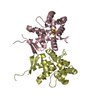
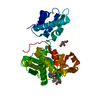
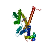
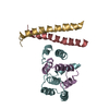
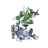
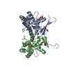
| Title | Crystal structure of a resolvase family site-specific recombinase from Streptococcus pneumoniae | ||||||
 Components Components | Site-specific recombinase, resolvase family protein | ||||||
 Keywords Keywords | RECOMBINATION / STRUCTURAL GENOMICS / RECOMBINASE / PSI-2 / Protein Structure Initiative / New York SGX Research Center for Structural Genomics / NYSGXRC / DNA BINDING PROTEIN | ||||||
| Function / homology | Resolvase, N-terminal catalytic domain / Rossmann fold / 3-Layer(aba) Sandwich / Alpha Beta / :  Function and homology information Function and homology information | ||||||
| Biological species |   Streptococcus pneumoniae (bacteria) Streptococcus pneumoniae (bacteria) | ||||||
| Method |  X-RAY DIFFRACTION / SYNCHROTRON / SAD / Resolution: 2.2 Å X-RAY DIFFRACTION / SYNCHROTRON / SAD / Resolution: 2.2 Å | ||||||
 Authors Authors | Bonanno, J.B. / Freeman, J. / Bain, K.T. / Do, J. / Sampathkumar, P. / Wasserman, S. / Sauder, J.M. / Burley, S.K. / Almo, S.C. / New York SGX Research Center for Structural Genomics (NYSGXRC) | ||||||
 Citation Citation | Journal: To be Published Title: Crystal structure of a resolvase family site-specific recombinase from Streptococcus pneumoniae Authors: Bonanno, J.B. / Freeman, J. / Bain, K.T. / Do, J. / Sampathkumar, P. / Wasserman, S. / Sauder, J.M. / Burley, S.K. / Almo, S.C. | ||||||
| History |
|
- Structure visualization
| Structure viewer | Molecule: MolmilJmol/JSmol |
|---|
- Downloads & links
-Download
| PDBx/mmCIF format | 3guv.cif.gz | 42.4 KB | Display | PDBx/mmCIF format |
|---|---|---|---|---|
| PDB format | pdb3guv.ent.gz | 29.2 KB | Display | PDB format |
| PDBx/mmJSON format | 3guv.json.gz | Tree view | PDBx/mmJSON format | |
| Others |  Other downloads Other downloads |
-Validation report
| Arichive directory | https://data.pdbj.org/pub/pdb/validation_reports/gu/3guvftp://data.pdbj.org/pub/pdb/validation_reports/gu/3guv | HTTPS FTP |
|---|
-Related structure data
| Similar structure data | |
|---|---|
| Other databases |


-Links
 PDBj
PDBj- Assembly
| Deposited unit | 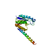
| ||||||||
|---|---|---|---|---|---|---|---|---|---|
| 1 | 
| ||||||||
| Unit cell |
| ||||||||
| Details | probable dimer |
-Components
| #1: Protein | Mass: 18878.541 Da / Num. of mol.: 1 Source method: isolated from a genetically manipulated source Source: (gene. exp.) Streptococcus pneumoniae (bacteria) / Strain: SP19-BS75 / Gene: CGSSp19BS75_09893 / Plasmid: modified pET26 / Production host: |
|---|---|
| #2: Water | ChemComp-HOH /  Mass: 18.015 Da / Num. of mol.: 30 / Source method: isolated from a natural source / Formula: H2O Mass: 18.015 Da / Num. of mol.: 30 / Source method: isolated from a natural source / Formula: H2O |
-Experimental details
-Experiment
| Experiment | Method: X-RAY DIFFRACTION / Number of used crystals: 1 |
|---|
- Sample preparation
| Crystal | Density Matthews: 2.01 Å3/Da / Density % sol: 38.91 % |
|---|---|
| Crystal grow | Temperature: 294 K / Method: vapor diffusion / pH: 7 Details: 20% PEG 3350, 200mM zinc acetate, pH 7.0, vapor diffusion, temperature 294K |
-Data collection
| Diffraction | Mean temperature: 100 K |
|---|---|
| Diffraction source | Source: SYNCHROTRON / Site: APS  / Beamline: 31-ID / Wavelength: 0.97958 Å / Beamline: 31-ID / Wavelength: 0.97958 Å |
| Detector | Type: MAR CCD 165 mm / Detector: CCD / Date: Mar 12, 2009 |
| Radiation | Monochromator: diamond / Protocol: SINGLE WAVELENGTH / Monochromatic (M) / Laue (L): M / Scattering type: x-ray |
| Radiation wavelength | Wavelength: 0.97958 Å / Relative weight: 1 |
| Reflection | Resolution: 2.2→38.519 Å / Num. all: 8257 / Num. obs: 8232 / % possible obs: 99.7 % / Observed criterion σ(F): 0 / Observed criterion σ(I): 0 / Redundancy: 5.9 % / Biso Wilson estimate: 34.8 Å2 / Rmerge(I) obs: 0.087 / Rsym value: 0.087 / Net I/σ(I): 14.1 |
| Reflection shell | Resolution: 2.2→2.32 Å / Redundancy: 6.1 % / Rmerge(I) obs: 0.225 / Mean I/σ(I) obs: 5.4 / Num. measured all: 7007 / Num. unique all: 1156 / Rsym value: 0.225 / % possible all: 100 |
- Processing
| Software |
| |||||||||||||||||||||||||||||||||||||||||||||||||||||||||||||||||||||||||||||||||||||
|---|---|---|---|---|---|---|---|---|---|---|---|---|---|---|---|---|---|---|---|---|---|---|---|---|---|---|---|---|---|---|---|---|---|---|---|---|---|---|---|---|---|---|---|---|---|---|---|---|---|---|---|---|---|---|---|---|---|---|---|---|---|---|---|---|---|---|---|---|---|---|---|---|---|---|---|---|---|---|---|---|---|---|---|---|---|---|
| Refinement | Method to determine structure: SAD / Resolution: 2.2→20 Å / Cor.coef. Fo:Fc: 0.939 / Cor.coef. Fo:Fc free: 0.907 / WRfactor Rfree: 0.336 / WRfactor Rwork: 0.973 / Occupancy max: 1 / Occupancy min: 0.5 / FOM work R set: 0.754 / SU B: 15.816 / SU ML: 0.18 / SU R Cruickshank DPI: 0.311 / SU Rfree: 0.253 / TLS residual ADP flag: LIKELY RESIDUAL / Cross valid method: THROUGHOUT / σ(F): 0 / σ(I): 0 / ESU R: 0.311 / ESU R Free: 0.253 / Stereochemistry target values: MAXIMUM LIKELIHOOD / Details: HYDROGENS HAVE BEEN ADDED IN THE RIDING POSITIONS
| |||||||||||||||||||||||||||||||||||||||||||||||||||||||||||||||||||||||||||||||||||||
| Solvent computation | Ion probe radii: 0.8 Å / Shrinkage radii: 0.8 Å / VDW probe radii: 1.4 Å / Solvent model: BABINET MODEL WITH MASK | |||||||||||||||||||||||||||||||||||||||||||||||||||||||||||||||||||||||||||||||||||||
| Displacement parameters | Biso max: 62.08 Å2 / Biso mean: 34.29 Å2 / Biso min: 13.36 Å2
| |||||||||||||||||||||||||||||||||||||||||||||||||||||||||||||||||||||||||||||||||||||
| Refinement step | Cycle: LAST / Resolution: 2.2→20 Å
| |||||||||||||||||||||||||||||||||||||||||||||||||||||||||||||||||||||||||||||||||||||
| Refine LS restraints |
| |||||||||||||||||||||||||||||||||||||||||||||||||||||||||||||||||||||||||||||||||||||
| LS refinement shell | Resolution: 2.2→2.257 Å / Total num. of bins used: 20
| |||||||||||||||||||||||||||||||||||||||||||||||||||||||||||||||||||||||||||||||||||||
| Refinement TLS params. | Method: refined / Refine-ID: X-RAY DIFFRACTION
| |||||||||||||||||||||||||||||||||||||||||||||||||||||||||||||||||||||||||||||||||||||
| Refinement TLS group |
|
