METAL BINDING PROTEIN / CBD1 / CBD2 / NCX / calcium binding domain 1 / Antiport / Calcium transport / Calmodulin-binding / Cell membrane / Glycoprotein / Ion transport / Membrane / Phosphoprotein / Sodium transport / Transmembrane / Transport / METAL TRANSPORT
Function / homology
Function and homology information
calcium:monoatomic cation antiporter activity involved in regulation of postsynaptic cytosolic calcium ion concentration / calcium:sodium antiporter activity / cell communication / sodium ion import across plasma membrane / intracellular sodium ion homeostasis / calcium ion import / ankyrin binding / calcium ion import across plasma membrane / positive regulation of the force of heart contraction / sodium ion transmembrane transport ...calcium:monoatomic cation antiporter activity involved in regulation of postsynaptic cytosolic calcium ion concentration / calcium:sodium antiporter activity / cell communication / sodium ion import across plasma membrane / intracellular sodium ion homeostasis / calcium ion import / ankyrin binding / calcium ion import across plasma membrane / positive regulation of the force of heart contraction / sodium ion transmembrane transport / positive regulation of bone mineralization / response to muscle stretch / calcium ion transmembrane transport / sarcolemma / postsynapse / calmodulin binding / axon / calcium ion binding / nucleoplasm / plasma membrane Similarity search - Function
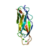
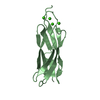
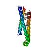
Sodium/calcium exchanger, isoform 1 / CalX-beta domain / Sodium/calcium exchanger protein / Sodium/calcium exchanger domain, C-terminal extension / C-terminal extension of sodium/calcium exchanger domain / Na-Ca exchanger/integrin-beta4 / Calx-beta domain / Domains in Na-Ca exchangers and integrin-beta4 / NCX, central ion-binding domain superfamily / Sodium/calcium exchanger membrane region ...Sodium/calcium exchanger, isoform 1 / CalX-beta domain / Sodium/calcium exchanger protein / Sodium/calcium exchanger domain, C-terminal extension / C-terminal extension of sodium/calcium exchanger domain / Na-Ca exchanger/integrin-beta4 / Calx-beta domain / Domains in Na-Ca exchangers and integrin-beta4 / NCX, central ion-binding domain superfamily / Sodium/calcium exchanger membrane region / Sodium/calcium exchanger protein / CalX-like domain superfamily / DnaJ domain / Immunoglobulin-like / Sandwich / Mainly Beta Similarity search - Domain/homology
In the structure databanks used in Yorodumi, some data are registered as the other names, "COVID-19 virus" and "2019-nCoV". Here are the details of the virus and the list of structure data.
Jan 31, 2019. EMDB accession codes are about to change! (news from PDBe EMDB page)
EMDB accession codes are about to change! (news from PDBe EMDB page)
The allocation of 4 digits for EMDB accession codes will soon come to an end. Whilst these codes will remain in use, new EMDB accession codes will include an additional digit and will expand incrementally as the available range of codes is exhausted. The current 4-digit format prefixed with “EMD-” (i.e. EMD-XXXX) will advance to a 5-digit format (i.e. EMD-XXXXX), and so on. It is currently estimated that the 4-digit codes will be depleted around Spring 2019, at which point the 5-digit format will come into force.
The EM Navigator/Yorodumi systems omit the EMD- prefix.
Related info.:Q: What is EMD? / ID/Accession-code notation in Yorodumi/EM Navigator
Yorodumi is a browser for structure data from EMDB, PDB, SASBDB, etc.
This page is also the successor to EM Navigator detail page, and also detail information page/front-end page for Omokage search.
The word "yorodu" (or yorozu) is an old Japanese word meaning "ten thousand". "mi" (miru) is to see.
Related info.:EMDB / PDB / SASBDB / Comparison of 3 databanks / Yorodumi Search / Aug 31, 2016. New EM Navigator & Yorodumi / Yorodumi Papers / Jmol/JSmol / Function and homology information / Changes in new EM Navigator and Yorodumi
 Movie
Movie Controller


 Open data
 Basic information
 Components
 Keywords
 Cell membrane /
  Function and homology information
 Authors
 Citation
 Structure visualization
 Downloads & links
 Other downloads


 PDBj
PDBj

 Assembly


 Mass: 40.078 Da / Num. of mol.: 7 / Source method: obtained synthetically / Formula: Ca
 Mass: 18.015 Da / Num. of mol.: 98 / Source method: isolated from a natural source / Formula: H2O
 Sample preparation
 / Beamline: 8.2.1 / Wavelength: 1 Å
 Processing