 Movie
Movie Controller

[English] 日本語
 Yorodumi
- PDB-3gbi: The Rational Design and Structural Analysis of a Self-Assembled T... -
+ Open data

- Basic information
| Entry | Database: PDB / ID: 3gbi | ||||||
|---|---|---|---|---|---|---|---|
| Title | The Rational Design and Structural Analysis of a Self-Assembled Three-Dimensional DNA Crystal | ||||||
 Components Components |
| ||||||
 Keywords Keywords |  DNA / nanotechnology / DNA crystals / DNA crossover / designed crystal lattice DNA / nanotechnology / DNA crystals / DNA crossover / designed crystal lattice | ||||||
| Function / homology | DNA / DNA (> 10) Function and homology information Function and homology information | ||||||
| Method | X-RAY DIFFRACTION / SYNCHROTRON / Resolution: 4.018 Å | ||||||
 Authors Authors | Birktoft, J.J. / Zheng, J. / Seeman, N.C. | ||||||
 Citation Citation | Journal: Nature / Year: 2009 Title: From molecular to macroscopic via the rational design of a self-assembled 3D DNA crystal. Authors: Zheng, J. / Birktoft, J.J. / Chen, Y. / Wang, T. / Sha, R. / Constantinou, P.E. / Ginell, S.L. / Mao, C. / Seeman, N.C. | ||||||
| History |
|
- Structure visualization
| Structure viewer | Molecule: MolmilJmol/JSmol |
|---|
- Downloads & links
-Download
| PDBx/mmCIF format | 3gbi.cif.gz | 30.2 KB | Display | PDBx/mmCIF format |
|---|---|---|---|---|
| PDB format | pdb3gbi.ent.gz | 19.8 KB | Display | PDB format |
| PDBx/mmJSON format | 3gbi.json.gz | Tree view | PDBx/mmJSON format | |
| Others |  Other downloads Other downloads |
-Validation report
| Arichive directory | https://data.pdbj.org/pub/pdb/validation_reports/gb/3gbiftp://data.pdbj.org/pub/pdb/validation_reports/gb/3gbi | HTTPS FTP |
|---|
-Related structure data
| Similar structure data |
|---|


-Links
 PDBj
PDBj


- Assembly
| Deposited unit | 
| ||||||||
|---|---|---|---|---|---|---|---|---|---|
| 1 | 
| ||||||||
| Unit cell |
| ||||||||
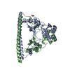
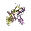
| Details | Authors state that the crystal is an infinite network made from three DNA strands that self-associate. In the current entry the asymmetric unit is comprised of 4 chains, 3 of which are fragments of longer DNA strands. Applying the space group H3 symmetry operators (X, Y, Z,), (-Y, X-Y, Z), and (-X+Y,-X,Z) to the contents of the asymmetric unit generates one trimeric unit of the self-associated DNA network. Additional details about the chemical composition and association are included in remark 400. |
-Components
| #1: DNA chain | Mass: 6457.188 Da / Num. of mol.: 1 / Source method: obtained synthetically Details: DNA strands were synthesized by standard phosphoramidite techniques on an Applied Biosystems 394 DNA synthesizer using trityl-on mode. |
|---|---|
| #2: DNA chain | Mass: 2082.400 Da / Num. of mol.: 1 Fragment: symmetrically- and sequentially repeating unit of a circular DNA molecules, see remark 400 for details. Source method: obtained synthetically Details: DNA strands were synthesized by standard phosphoramidite techniques on an Applied Biosystems 394 DNA synthesizer using trityl-on |
| #3: DNA chain | Mass: 1825.216 Da / Num. of mol.: 1 Fragment: last 6 residues of a DNA molecule used in experiment, see remark 400 for details. Source method: obtained synthetically Details: DNA strands were synthesized by standard phosphoramidite techniques on an Applied Biosystems 394 DNA synthesizer using trityl-on |
| #4: DNA chain | Mass: 2432.614 Da / Num. of mol.: 1 Fragment: first 8 residues of a DNA molecule used in experiment, see remark 400 for details. Source method: obtained synthetically Details: DNA strands were synthesized by standard phosphoramidite techniques on an Applied Biosystems 394 DNA synthesizer using trityl-on |
| Compound details | THE STRUCTURE UNIT IS GENERATED FROM 7 DNA STRANDS WHICH FORM A NETWORK UNIT WITH INTERNAL 3-FOLD ...THE STRUCTURE UNIT IS GENERATED FROM 7 DNA STRANDS WHICH FORM A NETWORK UNIT WITH INTERNAL 3-FOLD SYMMETRY. EXTENDING OF THE STRUCTURE UNIT IN 3-D SPACE RESULTS THE CRYSTAL AT R3 SPACE GROUP (REPRESENTE |
-Experimental details
-Experiment
| Experiment | Method: X-RAY DIFFRACTION / Number of used crystals: 1 |
|---|
- Sample preparation
| Crystal grow | Temperature: 293 K / Method: vapor diffusion, sitting drop / pH: 8.5 Details: Grown by vapor diffusion while treated with a controlled temperature gradient from 333 degs to 293 degs., pH 8.5, VAPOR DIFFUSION, SITTING DROP |
|---|
-Data collection
| Diffraction source | Source: SYNCHROTRON / Site: APS  / Beamline: 19-ID / Wavelength: 1 Å / Beamline: 19-ID / Wavelength: 1 Å |
|---|---|
| Detector | Type: ADSC QUANTUM 315r / Detector: CCD / Date: Jun 17, 2008 |
| Radiation | Monochromator: Graphite / Protocol: SINGLE WAVELENGTH / Monochromatic (M) / Laue (L): M / Scattering type: x-ray |
| Radiation wavelength | Wavelength: 1 Å / Relative weight: 1 |
| Reflection | Resolution: 4.01→17.3 Å / Num. all: 3341 / Num. obs: 3211 / % possible obs: 96.1 % / Observed criterion σ(F): 0 / Observed criterion σ(I): 0 / Redundancy: 7.3 % / Biso Wilson estimate: 199.88 Å2 / Rmerge(I) obs: 0.067 / Net I/σ(I): 10.9 |
| Reflection shell | Resolution: 4→4.07 Å / % possible all: 100 |
- Processing
| Software |
| ||||||||||||||||||||||||||||
|---|---|---|---|---|---|---|---|---|---|---|---|---|---|---|---|---|---|---|---|---|---|---|---|---|---|---|---|---|---|
| Refinement | Resolution: 4.018→17.288 Å / Occupancy max: 1 / Occupancy min: 1 / SU ML: 0.83 / σ(F): 0.14 / Stereochemistry target values: ML Details: In reality all 3211 measured reflections were used in the refinement (using PHENIX). Of these only 2022 had FOBS/SIGMA_FOBS >/= 0.140.
| ||||||||||||||||||||||||||||
| Solvent computation | Solvent model: FLAT BULK SOLVENT MODEL / Bsol: 20.001 Å2 / ksol: 0.09 e/Å3 | ||||||||||||||||||||||||||||
| Displacement parameters | Biso mean: 80 Å2
| ||||||||||||||||||||||||||||
| Refinement step | Cycle: LAST / Resolution: 4.018→17.288 Å
| ||||||||||||||||||||||||||||
| Refine LS restraints |
| ||||||||||||||||||||||||||||
| LS refinement shell | Highest resolution: 4.018 Å / Total num. of bins used: 1
|
