 Movie
Movie Controller

[English] 日本語
 Yorodumi
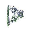
- PDB-5w6w: Designing Higher Resolution Self-Assembled 3D DNA Crystals via St... -
+ Open data

- Basic information
| Entry | Database: PDB / ID: 5w6w | ||||||
|---|---|---|---|---|---|---|---|
| Title | Designing Higher Resolution Self-Assembled 3D DNA Crystals via Strand Terminus Modifications | ||||||
 Components Components |
| ||||||
 Keywords Keywords | DNA / nanotechnology / DNA crystals / DNA lattices | ||||||
| Function / homology | DNA / DNA (> 10) Function and homology information Function and homology information | ||||||
| Biological species | synthetic construct (others) | ||||||
| Method |  X-RAY DIFFRACTION / SYNCHROTRON / MOLECULAR REPLACEMENT / molecular replacement / Resolution: 3.06 Å X-RAY DIFFRACTION / SYNCHROTRON / MOLECULAR REPLACEMENT / molecular replacement / Resolution: 3.06 Å | ||||||
 Authors Authors | Hernandez, C. / Birktoft, J.J. / Seeman, N.C. | ||||||
| Funding support |  United States, 1items United States, 1items
| ||||||
 Citation Citation | Journal: To Be Published Title: Designing Higher Resolution Self-Assembled 3D DNA Crystals via Strand Terminus Modifications Authors: Hernandez, C. / Birktoft, J.J. / Seeman, N.C. / Ohayon, Y.P. / Chandrasekaran, A.R. / Abdalla, H.O. / Mohsen, M. / Sha, R. / Chenge, M. / Lukeman, P.S. / Chaikin, P. / Ginell, S.L. / Jong, M.A. | ||||||
| History |
|
- Structure visualization
| Structure viewer | Molecule: MolmilJmol/JSmol |
|---|
- Downloads & links
-Download
| PDBx/mmCIF format | 5w6w.cif.gz | 32.9 KB | Display | PDBx/mmCIF format |
|---|---|---|---|---|
| PDB format | pdb5w6w.ent.gz | 22.5 KB | Display | PDB format |
| PDBx/mmJSON format | 5w6w.json.gz | Tree view | PDBx/mmJSON format | |
| Others |  Other downloads Other downloads |
-Validation report
| Arichive directory | https://data.pdbj.org/pub/pdb/validation_reports/w6/5w6wftp://data.pdbj.org/pub/pdb/validation_reports/w6/5w6w | HTTPS FTP |
|---|
-Related structure data
| Related structure data | 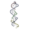 3gbiS S: Starting model for refinement |
|---|---|
| Similar structure data |


-Links
 PDBj
PDBj


- Assembly
| Deposited unit | 
| ||||||||
|---|---|---|---|---|---|---|---|---|---|
| 1 | 
| ||||||||
| Unit cell |
|
-Components
| #1: DNA chain | Mass: 6457.188 Da / Num. of mol.: 1 / Source method: obtained synthetically / Source: (synth.) synthetic construct (others) |
|---|---|
| #2: DNA chain | Mass: 2082.400 Da / Num. of mol.: 1 / Source method: obtained synthetically / Source: (synth.) synthetic construct (others) |
| #3: DNA chain | Mass: 2129.409 Da / Num. of mol.: 1 / Source method: obtained synthetically / Source: (synth.) synthetic construct (others) |
| #4: DNA chain | Mass: 2128.421 Da / Num. of mol.: 1 / Source method: obtained synthetically / Source: (synth.) synthetic construct (others) |
-Experimental details
-Experiment
| Experiment | Method: X-RAY DIFFRACTION / Number of used crystals: 1 |
|---|
- Sample preparation
| Crystal | Density Matthews: 3.75 Å3/Da / Density % sol: 67 % / Description: rhombohedral |
|---|---|
| Crystal grow | Temperature: 293 K / Method: vapor diffusion, hanging drop / pH: 7 Details: Grown by vapor diffusion while treated with a controlled temperature gradient from 333K degs to 293K. They were grown in Ammonium Sulfate, TRIS, Acetic Acid and EDTA. |
-Data collection
| Diffraction | Mean temperature: 100 K |
|---|---|
| Diffraction source | Source: SYNCHROTRON / Site: APS / Beamline: 19-ID / Wavelength: 1.1 Å |
| Detector | Type: ADSC QUANTUM 315 / Detector: CCD / Date: Oct 14, 2013 |
| Radiation | Protocol: SINGLE WAVELENGTH / Monochromatic (M) / Laue (L): M / Scattering type: x-ray |
| Radiation wavelength | Wavelength: 1.1 Å / Relative weight: 1 |
| Reflection | Resolution: 3.05→50 Å / Num. obs: 5152 / % possible obs: 68 % / Redundancy: 7.8 % / Biso Wilson estimate: 90.04 Å2 / Rmerge(I) obs: 0.107 / Net I/σ(I): 10.8 |
| Reflection shell | Resolution: 3.05→3.1 Å / Redundancy: 6.5 % / Rmerge(I) obs: 0.431 / % possible all: 19.2 |
-Phasing
| Phasing | Method: molecular replacement |
|---|
- Processing
| Software |
| |||||||||||||||||||||||||||||||||||
|---|---|---|---|---|---|---|---|---|---|---|---|---|---|---|---|---|---|---|---|---|---|---|---|---|---|---|---|---|---|---|---|---|---|---|---|---|
| Refinement | Method to determine structure: MOLECULAR REPLACEMENT Starting model: 3GBI Resolution: 3.06→32.96 Å / SU ML: 0.39 / Cross valid method: FREE R-VALUE / σ(F): 1.96 / Phase error: 50.68 / Stereochemistry target values: ML
| |||||||||||||||||||||||||||||||||||
| Solvent computation | Shrinkage radii: 0.9 Å / VDW probe radii: 1.11 Å / Solvent model: FLAT BULK SOLVENT MODEL | |||||||||||||||||||||||||||||||||||
| Displacement parameters | Biso mean: 120.6 Å2 | |||||||||||||||||||||||||||||||||||
| Refinement step | Cycle: LAST / Resolution: 3.06→32.96 Å
| |||||||||||||||||||||||||||||||||||
| Refine LS restraints |
| |||||||||||||||||||||||||||||||||||
| LS refinement shell |
|
