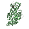
 Movie
Movie Controller


+ Open data

- Basic information
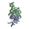
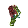
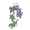
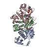
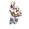
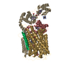
| Entry | Database: PDB / ID: 3ez9 | ||||||
|---|---|---|---|---|---|---|---|
| Title | Partition Protein | ||||||
 Components Components | ParA | ||||||
 Keywords Keywords |  BIOSYNTHETIC PROTEIN / dna binding / winged-hth / partition BIOSYNTHETIC PROTEIN / dna binding / winged-hth / partition | ||||||
| Function / homology | Multidrug-efflux Transporter Regulator; Chain: A; Domain 2 - #30 / Multidrug-efflux Transporter Regulator; Chain: A; Domain 2 / P-loop containing nucleotide triphosphate hydrolases / Rossmann fold / Orthogonal Bundle / 3-Layer(aba) Sandwich / Mainly Alpha / Alpha Beta / :  Function and homology information Function and homology information | ||||||
| Biological species |  Salmonella enterica subsp. enterica serovar Newport str. SL317 (bacteria) Salmonella enterica subsp. enterica serovar Newport str. SL317 (bacteria) | ||||||
| Method | X-RAY DIFFRACTION / SYNCHROTRON / Resolution: 2.8 Å | ||||||
 Authors Authors | Schumacher, M.A. | ||||||
 Citation Citation | Journal: Embo J. / Year: 2009 Title: Structural basis for ADP-mediated transcriptional regulation by P1 and P7 ParA. Authors: Dunham, T.D. / Xu, W. / Funnell, B.E. / Schumacher, M.A. | ||||||
| History |
|
- Structure visualization
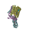
| Structure viewer | Molecule: MolmilJmol/JSmol |
|---|
- Downloads & links
-Download
| PDBx/mmCIF format | 3ez9.cif.gz | 152.3 KB | Display | PDBx/mmCIF format |
|---|---|---|---|---|
| PDB format | pdb3ez9.ent.gz | 121.3 KB | Display | PDB format |
| PDBx/mmJSON format | 3ez9.json.gz | Tree view | PDBx/mmJSON format | |
| Others |  Other downloads Other downloads |
-Validation report
| Arichive directory | https://data.pdbj.org/pub/pdb/validation_reports/ez/3ez9ftp://data.pdbj.org/pub/pdb/validation_reports/ez/3ez9 | HTTPS FTP |
|---|
-Related structure data


-Links
 PDBj
PDBj- Assembly
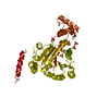
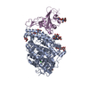
| Deposited unit | 
| ||||||||
|---|---|---|---|---|---|---|---|---|---|
| 1 | 
| ||||||||
| 2 | 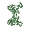
| ||||||||
| Unit cell |
| ||||||||
| Details | dimer |
-Components
| #1: Protein | Mass: 45534.625 Da / Num. of mol.: 2 Source method: isolated from a genetically manipulated source Source: (gene. exp.) Salmonella enterica subsp. enterica serovar Newport str. SL317 (bacteria)Gene: SNSL317_A0932 / References: UniProt: B4ABW6 #2: Chemical | ChemComp-MG / |   Mass: 24.305 Da / Num. of mol.: 1 / Source method: obtained synthetically / Formula: Mg Mass: 24.305 Da / Num. of mol.: 1 / Source method: obtained synthetically / Formula: Mg#3: Water | ChemComp-HOH / | Water Mass: 18.015 Da / Num. of mol.: 75 / Source method: isolated from a natural source / Formula: H2O Mass: 18.015 Da / Num. of mol.: 75 / Source method: isolated from a natural source / Formula: H2O |
|---|
-Experimental details
-Experiment
| Experiment | Method: X-RAY DIFFRACTION / Number of used crystals: 1 |
|---|
- Sample preparation
| Crystal | Density Matthews: 3.18 Å3/Da / Density % sol: 61.28 % |
|---|---|
| Crystal grow | Temperature: 298 K / Method: vapor diffusion, hanging drop / pH: 7.5 Details: Peg 3000, ethylene glycol, pH 7.5, VAPOR DIFFUSION, HANGING DROP, temperature 298K |
-Data collection
| Diffraction | Mean temperature: 100 K |
|---|---|
| Diffraction source | Source: SYNCHROTRON / Site: ALS  / Beamline: 8.2.1 / Wavelength: 1 Å / Beamline: 8.2.1 / Wavelength: 1 Å |
| Detector | Type: ADSC QUANTUM 4 / Detector: CCD / Date: Jan 2, 2007 / Details: mirrors |
| Radiation | Monochromator: mirrors / Protocol: SINGLE WAVELENGTH / Monochromatic (M) / Laue (L): M / Scattering type: x-ray |
| Radiation wavelength | Wavelength: 1 Å / Relative weight: 1 |
| Reflection | Resolution: 2.78→100.5 Å / Num. obs: 33374 / % possible obs: 96 % / Observed criterion σ(F): 0 / Observed criterion σ(I): 0 / Redundancy: 3 % / Biso Wilson estimate: 52.7 Å2 / Rsym value: 0.048 / Net I/σ(I): 10.1 |
- Processing
| Software | Name: CNS / Version: 1.2 / Classification: refinement | |||||||||||||||||||||||||||
|---|---|---|---|---|---|---|---|---|---|---|---|---|---|---|---|---|---|---|---|---|---|---|---|---|---|---|---|---|
| Refinement | Resolution: 2.8→66.85 Å / Rfactor Rfree error: 0.007 / Occupancy max: 1 / Occupancy min: 0.5 / Data cutoff high absF: 2254383 / Data cutoff low absF: 0 / Isotropic thermal model: RESTRAINED / Cross valid method: THROUGHOUT / σ(F): 0 / Details: BULK SOLVENT MODEL USED
| |||||||||||||||||||||||||||
| Solvent computation | Solvent model: FLAT MODEL / Bsol: 58.177 Å2 / ksol: 0.3 e/Å3 | |||||||||||||||||||||||||||
| Displacement parameters | Biso max: 184.8 Å2 / Biso mean: 87.502 Å2 / Biso min: 20 Å2
| |||||||||||||||||||||||||||
| Refine analyze |
| |||||||||||||||||||||||||||
| Refinement step | Cycle: LAST / Resolution: 2.8→66.85 Å
| |||||||||||||||||||||||||||
| Refine LS restraints |
| |||||||||||||||||||||||||||
| LS refinement shell | Resolution: 2.8→2.98 Å / Rfactor Rfree error: 0.023 / Total num. of bins used: 6
| |||||||||||||||||||||||||||
| Xplor file |
|
