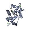
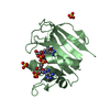
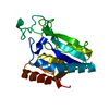
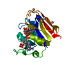
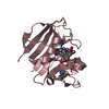
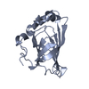
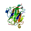
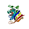
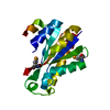
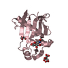
[English] 日本語
Yorodumi
- PDB-3ewt: Crystal Structure of calmodulin complexed with a peptide -

+
Open data


ID or keywords:

Loading...

-
Basic information

Entry
Database: PDB / ID: 3ewt
TitleCrystal Structure of calmodulin complexed with a peptide
Components
  • Calmodulin
  • Tumor necrosis factor receptor superfamily member 6
KeywordsCALCIUM BINDING PROTEIN / Calmodulin-peptide complex / Fas / death domain
Function / homology
Function and homology information


Fas signaling pathway / cellular response to hyperoxia / FasL/ CD95L signaling / tumor necrosis factor receptor activity / CD95 death-inducing signaling complex / activation-induced cell death of T cells / positive regulation of cysteine-type endopeptidase activity involved in apoptotic signaling pathway / Regulation by c-FLIP / CASP8 activity is inhibited / Dimerization of procaspase-8 ...Fas signaling pathway / cellular response to hyperoxia / FasL/ CD95L signaling / tumor necrosis factor receptor activity / CD95 death-inducing signaling complex / activation-induced cell death of T cells / positive regulation of cysteine-type endopeptidase activity involved in apoptotic signaling pathway / Regulation by c-FLIP / CASP8 activity is inhibited / Dimerization of procaspase-8 / Caspase activation via Death Receptors in the presence of ligand / : / necroptotic signaling pathway / establishment of protein localization to mitochondrial membrane / type 3 metabotropic glutamate receptor binding / death-inducing signaling complex / CaM pathway / Cam-PDE 1 activation / Sodium/Calcium exchangers / motor neuron apoptotic process / Calmodulin induced events / TP53 Regulates Transcription of Death Receptors and Ligands / Reduction of cytosolic Ca++ levels / Activation of Ca-permeable Kainate Receptor / CREB1 phosphorylation through the activation of CaMKII/CaMKK/CaMKIV cascasde / regulation of synaptic vesicle endocytosis / Loss of phosphorylation of MECP2 at T308 / RIPK1-mediated regulated necrosis / CREB1 phosphorylation through the activation of Adenylate Cyclase / PKA activation / negative regulation of high voltage-gated calcium channel activity / regulation of synaptic vesicle exocytosis / CaMK IV-mediated phosphorylation of CREB / Glycogen breakdown (glycogenolysis) / negative regulation of calcium ion export across plasma membrane / organelle localization by membrane tethering / Activation of RAC1 downstream of NMDARs / regulation of cardiac muscle cell action potential / mitochondrion-endoplasmic reticulum membrane tethering / CLEC7A (Dectin-1) induces NFAT activation / regulation of stress-activated MAPK cascade / autophagosome membrane docking / response to corticosterone / positive regulation of ryanodine-sensitive calcium-release channel activity / Negative regulation of NMDA receptor-mediated neuronal transmission / nitric-oxide synthase binding / regulation of cell communication by electrical coupling involved in cardiac conduction / Unblocking of NMDA receptors, glutamate binding and activation / negative regulation of peptidyl-threonine phosphorylation / Synthesis of IP3 and IP4 in the cytosol / Phase 0 - rapid depolarisation / protein phosphatase activator activity / RHO GTPases activate PAKs / positive regulation of cyclic-nucleotide phosphodiesterase activity / positive regulation of phosphoprotein phosphatase activity / Long-term potentiation / Ion transport by P-type ATPases / Uptake and function of anthrax toxins / adenylate cyclase binding / Calcineurin activates NFAT / Regulation of MECP2 expression and activity / catalytic complex / DARPP-32 events / detection of calcium ion / negative regulation of ryanodine-sensitive calcium-release channel activity / Smooth Muscle Contraction / RHO GTPases activate IQGAPs / regulation of cardiac muscle contraction / calcium channel inhibitor activity / cellular response to interferon-beta / positive regulation of DNA binding / regulation of cardiac muscle contraction by regulation of the release of sequestered calcium ion / Protein methylation / enzyme regulator activity / voltage-gated potassium channel complex / phosphatidylinositol 3-kinase binding / Activation of AMPK downstream of NMDARs / eNOS activation / regulation of release of sequestered calcium ion into cytosol by sarcoplasmic reticulum / positive regulation of apoptotic signaling pathway / extrinsic apoptotic signaling pathway / regulation of calcium-mediated signaling / positive regulation of protein dephosphorylation / titin binding / regulation of ryanodine-sensitive calcium-release channel activity / Tetrahydrobiopterin (BH4) synthesis, recycling, salvage and regulation / Ion homeostasis / positive regulation of protein autophosphorylation / extrinsic apoptotic signaling pathway in absence of ligand / sperm midpiece / response to amphetamine / calcium channel complex / cellular response to amino acid starvation / activation of adenylate cyclase activity / substantia nigra development / adenylate cyclase activator activity / Ras activation upon Ca2+ influx through NMDA receptor / regulation of heart rate / nitric-oxide synthase regulator activity / protein serine/threonine kinase activator activity
Similarity search - Function
Fas receptor / Fas receptor, death domain / Fas receptor, N-terminal / TNFR/NGFR family cysteine-rich region domain profile. / TNFR/NGFR cysteine-rich region / TNFR/NGFR family cysteine-rich region signature. / Tumor necrosis factor receptor / nerve growth factor receptor repeats. / TNFR/NGFR cysteine-rich region / Death domain profile. / DEATH domain, found in proteins involved in cell death (apoptosis). ...Fas receptor / Fas receptor, death domain / Fas receptor, N-terminal / TNFR/NGFR family cysteine-rich region domain profile. / TNFR/NGFR cysteine-rich region / TNFR/NGFR family cysteine-rich region signature. / Tumor necrosis factor receptor / nerve growth factor receptor repeats. / TNFR/NGFR cysteine-rich region / Death domain profile. / DEATH domain, found in proteins involved in cell death (apoptosis). / Death domain / Death domain / Death-like domain superfamily / EF-hand domain pair / EF-hand, calcium binding motif / EF-Hand 1, calcium-binding site / EF-hand calcium-binding domain. / EF-hand calcium-binding domain profile. / EF-hand domain / EF-hand domain pair
Similarity search - Domain/homology
Calmodulin-1 / Tumor necrosis factor receptor superfamily member 6 / Calmodulin-3
Similarity search - Component
Biological speciesHomo sapiens (human)
synthetic construct (others)
MethodX-RAY DIFFRACTION / MOLECULAR REPLACEMENT / Resolution: 2.4 Å
AuthorsJiang, T. / Cao, P. / Gong, Y. / Yu, H.J. / Gui, W.J. / Zhang, W.T.
CitationJournal: Acta Crystallogr.,Sect.D / Year: 2014
Title: Structural insights into the mechanism of calmodulin binding to death receptors.
Authors: Cao, P. / Zhang, W. / Gui, W. / Dong, Y. / Jiang, T. / Gong, Y.
History
DepositionOct 16, 2008Deposition site: RCSB / Processing site: PDBJ
Revision 1.0Oct 20, 2009Provider: repository / Type: Initial release
Revision 1.1Jul 13, 2011Group: Version format compliance
Revision 1.2Jun 18, 2014Group: Database references
Revision 1.3Jan 1, 2020Group: Database references / Source and taxonomy
Category: citation / citation_author ...citation / citation_author / pdbx_entity_src_syn / struct_ref_seq_dif
Item: _citation.country / _citation.journal_id_CSD ..._citation.country / _citation.journal_id_CSD / _citation.journal_id_ISSN / _citation.pdbx_database_id_PubMed / _citation.title / _citation_author.name / _pdbx_entity_src_syn.ncbi_taxonomy_id / _pdbx_entity_src_syn.organism_scientific / _struct_ref_seq_dif.details
Revision 1.4Nov 1, 2023Group: Data collection / Database references ...Data collection / Database references / Derived calculations / Refinement description
Category: chem_comp_atom / chem_comp_bond ...chem_comp_atom / chem_comp_bond / database_2 / pdbx_initial_refinement_model / struct_site
Item: _database_2.pdbx_DOI / _database_2.pdbx_database_accession ..._database_2.pdbx_DOI / _database_2.pdbx_database_accession / _struct_site.pdbx_auth_asym_id / _struct_site.pdbx_auth_comp_id / _struct_site.pdbx_auth_seq_id

-
Structure visualization

Structure viewerMolecule:
MolmilJmol/JSmol

Downloads & links

-
Assembly

Deposited unit
A: Calmodulin
E: Tumor necrosis factor receptor superfamily member 6
hetero molecules


Theoretical massNumber of molelcules
Total (without water)20,4136
Polymers20,2522
Non-polymers1604
Water79344
1


  • Idetical with deposited unit
  • defined by author&software
TypeNameSymmetry operationNumber
identity operation1_555x,y,z1
Buried area2620 Å2
ΔGint-78 kcal/mol
Surface area8310 Å2
MethodPISA
Unit cell
Length a, b, c (Å)40.062, 40.062, 174.431
Angle α, β, γ (deg.)90.00, 90.00, 120.00
Int Tables number154
Space group name H-MP3221
Components on special symmetry positions
IDModelComponents
11A-626-

HOH

21A-627-

HOH

-
Components

#1: Protein Calmodulin / / CaM


Mass: 17550.232 Da / Num. of mol.: 1
Source method: isolated from a genetically manipulated source
Source: (gene. exp.) Homo sapiens (human) / Plasmid: pET28a / Production host: Escherichia coli BL21(DE3) (bacteria) / Strain (production host): BL21(DE3) / References: UniProt: P62158, UniProt: P0DP23*PLUS
#2: Protein/peptide Tumor necrosis factor receptor superfamily member 6 / FASLG receptor / Apoptosis-mediating surface antigen FAS / Apo-1 antigen


Mass: 2702.220 Da / Num. of mol.: 1 / Fragment: Helix(1+2) of death domain / Source method: obtained synthetically / Details: This sequence occurs naturally in humans. / Source: (synth.) synthetic construct (others) / References: UniProt: P25445
#3: Chemical
ChemComp-CA / CALCIUM ION


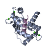
Mass: 40.078 Da / Num. of mol.: 4 / Source method: obtained synthetically / Formula: Ca
#4: Water ChemComp-HOH / water / Water


Mass: 18.015 Da / Num. of mol.: 44 / Source method: isolated from a natural source / Formula: H2O

-
Experimental details

-
Experiment

ExperimentMethod: X-RAY DIFFRACTION / Number of used crystals: 1

-
Sample preparation

CrystalDensity Matthews: 2 Å3/Da / Density % sol: 38.35 %
Crystal growTemperature: 293 K / Method: evaporation / pH: 5.5
Details: 25%(w/v) PEG8000, 0.2M Sodium Acetate, 0.1M Sodium cacodylate, pH 5.5, EVAPORATION, temperature 293K

-
Data collection

DiffractionMean temperature: 113 K
Diffraction sourceSource: ROTATING ANODE / Type: RIGAKU / Wavelength: 1.5418 Å
DetectorType: RIGAKU RAXIS IV++ / Detector: IMAGE PLATE / Date: Sep 22, 2006
RadiationMonochromator: GRAPHITE / Protocol: SINGLE WAVELENGTH / Monochromatic (M) / Laue (L): M / Scattering type: x-ray
Radiation wavelengthWavelength: 1.5418 Å / Relative weight: 1
ReflectionResolution: 2.4→20 Å / Num. obs: 6946 / % possible obs: 99.6 % / Observed criterion σ(F): 2 / Observed criterion σ(I): 2 / Redundancy: 8.6 % / Rmerge(I) obs: 0.042 / Rsym value: 0.041
Reflection shellResolution: 2.4→2.46 Å / % possible all: 97.3

-
Processing

Software
NameVersionClassification
CrystalCleardata collection
MOLREPphasing
REFMAC5.2.0019refinement
DENZOdata reduction
SCALEPACKdata scaling
RefinementMethod to determine structure: MOLECULAR REPLACEMENT
Starting model: 1L7Z
Resolution: 2.4→19.9 Å / Cor.coef. Fo:Fc: 0.929 / Cor.coef. Fo:Fc free: 0.901 / Cross valid method: THROUGHOUT / σ(F): 0 / σ(I): 0 / ESU R: 0.491 / ESU R Free: 0.278 / Stereochemistry target values: MAXIMUM LIKELIHOOD / Details: HYDROGENS HAVE BEEN ADDED IN THE RIDING POSITIONS
RfactorNum. reflection% reflectionSelection details
Rfree0.25916 312 4.5 %RANDOM
Rwork0.21664 ---
all0.21859 6575 --
obs0.21859 6575 99.8 %-
Solvent computationIon probe radii: 0.8 Å / Shrinkage radii: 0.8 Å / VDW probe radii: 1.2 Å / Solvent model: MASK
Displacement parametersBiso mean: 35.709 Å2
Baniso -1Baniso -2Baniso -3
1-0.73 Å20.36 Å20 Å2
2--0.73 Å20 Å2
3----1.09 Å2
Refinement stepCycle: LAST / Resolution: 2.4→19.9 Å
ProteinNucleic acidLigandSolventTotal
Num. atoms1227 0 4 44 1275
Refine LS restraints
Refine-IDTypeDev idealDev ideal targetNumber
X-RAY DIFFRACTIONr_bond_refined_d0.0110.0221239
X-RAY DIFFRACTIONr_bond_other_d
X-RAY DIFFRACTIONr_angle_refined_deg1.3841.9631667
X-RAY DIFFRACTIONr_angle_other_deg
X-RAY DIFFRACTIONr_dihedral_angle_1_deg5.2935157
X-RAY DIFFRACTIONr_dihedral_angle_2_deg37.66126.30865
X-RAY DIFFRACTIONr_dihedral_angle_3_deg20.23815224
X-RAY DIFFRACTIONr_dihedral_angle_4_deg16.376156
X-RAY DIFFRACTIONr_chiral_restr0.110.2189
X-RAY DIFFRACTIONr_gen_planes_refined0.0040.02940
X-RAY DIFFRACTIONr_gen_planes_other
X-RAY DIFFRACTIONr_nbd_refined0.240.2682
X-RAY DIFFRACTIONr_nbd_other
X-RAY DIFFRACTIONr_nbtor_refined0.3010.2880
X-RAY DIFFRACTIONr_nbtor_other
X-RAY DIFFRACTIONr_xyhbond_nbd_refined0.1280.257
X-RAY DIFFRACTIONr_xyhbond_nbd_other
X-RAY DIFFRACTIONr_metal_ion_refined0.0970.221
X-RAY DIFFRACTIONr_metal_ion_other
X-RAY DIFFRACTIONr_symmetry_vdw_refined0.2680.232
X-RAY DIFFRACTIONr_symmetry_vdw_other
X-RAY DIFFRACTIONr_symmetry_hbond_refined0.1850.26
X-RAY DIFFRACTIONr_symmetry_hbond_other
X-RAY DIFFRACTIONr_symmetry_metal_ion_refined
X-RAY DIFFRACTIONr_symmetry_metal_ion_other
X-RAY DIFFRACTIONr_mcbond_it0.9161.5783
X-RAY DIFFRACTIONr_mcbond_other
X-RAY DIFFRACTIONr_mcangle_it1.49921252
X-RAY DIFFRACTIONr_scbond_it1.73456
X-RAY DIFFRACTIONr_scangle_it3.0234.5415
X-RAY DIFFRACTIONr_rigid_bond_restr
X-RAY DIFFRACTIONr_sphericity_free
X-RAY DIFFRACTIONr_sphericity_bonded
LS refinement shellResolution: 2.401→2.463 Å / Total num. of bins used: 20
RfactorNum. reflection% reflection
Rfree0.447 18 -
Rwork0.249 475 -
obs--99.6 %

+
About Yorodumi

-
News

-
Feb 9, 2022. New format data for meta-information of EMDB entries

New format data for meta-information of EMDB entries

  • Version 3 of the EMDB header file is now the official format.
  • The previous official version 1.9 will be removed from the archive.

Related info.:EMDB header

External links:wwPDB to switch to version 3 of the EMDB data model

-
Aug 12, 2020. Covid-19 info

Covid-19 info

URL: https://pdbj.org/emnavi/covid19.php

New page: Covid-19 featured information page in EM Navigator.

Related info.:Covid-19 info / Mar 5, 2020. Novel coronavirus structure data

+
Mar 5, 2020. Novel coronavirus structure data

Novel coronavirus structure data

Related info.:Yorodumi Speices / Aug 12, 2020. Covid-19 info

External links:COVID-19 featured content - PDBj / Molecule of the Month (242):Coronavirus Proteases

+
Jan 31, 2019. EMDB accession codes are about to change! (news from PDBe EMDB page)

EMDB accession codes are about to change! (news from PDBe EMDB page)

  • The allocation of 4 digits for EMDB accession codes will soon come to an end. Whilst these codes will remain in use, new EMDB accession codes will include an additional digit and will expand incrementally as the available range of codes is exhausted. The current 4-digit format prefixed with “EMD-” (i.e. EMD-XXXX) will advance to a 5-digit format (i.e. EMD-XXXXX), and so on. It is currently estimated that the 4-digit codes will be depleted around Spring 2019, at which point the 5-digit format will come into force.
  • The EM Navigator/Yorodumi systems omit the EMD- prefix.

Related info.:Q: What is EMD? / ID/Accession-code notation in Yorodumi/EM Navigator

External links:EMDB Accession Codes are Changing Soon! / Contact to PDBj

+
Jul 12, 2017. Major update of PDB

Major update of PDB

  • wwPDB released updated PDB data conforming to the new PDBx/mmCIF dictionary.
  • This is a major update changing the version number from 4 to 5, and with Remediation, in which all the entries are updated.
  • In this update, many items about electron microscopy experimental information are reorganized (e.g. em_software).
  • Now, EM Navigator and Yorodumi are based on the updated data.

External links:wwPDB Remediation / Enriched Model Files Conforming to OneDep Data Standards Now Available in the PDB FTP Archive

-
Yorodumi

Thousand views of thousand structures

  • Yorodumi is a browser for structure data from EMDB, PDB, SASBDB, etc.
  • This page is also the successor to EM Navigator detail page, and also detail information page/front-end page for Omokage search.
  • The word "yorodu" (or yorozu) is an old Japanese word meaning "ten thousand". "mi" (miru) is to see.

Related info.:EMDB / PDB / SASBDB / Comparison of 3 databanks / Yorodumi Search / Aug 31, 2016. New EM Navigator & Yorodumi / Yorodumi Papers / Jmol/JSmol / Function and homology information / Changes in new EM Navigator and Yorodumi

Read more