 Movie
Movie Controller

[English] 日本語
 Yorodumi
- PDB-1ilw: Crystal Structure of Pyrazinamidase/Nicotinamidase of Pyrococcus ... -
+ Open data

- Basic information
| Entry | Database: PDB / ID: 1ilw | ||||||
|---|---|---|---|---|---|---|---|
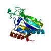
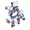
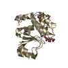
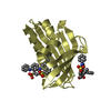
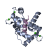
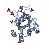
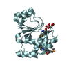
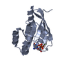
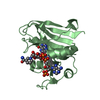
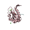
| Title | Crystal Structure of Pyrazinamidase/Nicotinamidase of Pyrococcus horikoshii | ||||||
 Components Components | 180 aa long hypothetical Pyrazinamidase/nicotinamidase | ||||||
 Keywords Keywords | HYDROLASE / Pyrazinamide / pyrazinamidase / nicotinamidase / tuberculosis / cysteine hydrolase / amidase / Structural Genomics / BSGC structure funded by NIH / Protein Structure Initiative / PSI / Berkeley Structural Genomics Center | ||||||
| Function / homology |  Function and homology information Function and homology informationpyridine nucleotide biosynthetic process / nicotinamidase / nicotinamidase activity / metal ion binding Similarity search - Function | ||||||
| Biological species |   Pyrococcus horikoshii (archaea) Pyrococcus horikoshii (archaea) | ||||||
| Method |  X-RAY DIFFRACTION / SYNCHROTRON / MOLECULAR REPLACEMENT / Resolution: 2.05 Å X-RAY DIFFRACTION / SYNCHROTRON / MOLECULAR REPLACEMENT / Resolution: 2.05 Å | ||||||
 Authors Authors | Du, X. / Kim, S.-H. / Berkeley Structural Genomics Center (BSGC) | ||||||
 Citation Citation | Journal: Biochemistry / Year: 2001 Title: Crystal structure and mechanism of catalysis of a pyrazinamidase from Pyrococcus horikoshii. Authors: Du, X. / Wang, W. / Kim, R. / Yakota, H. / Nguyen, H. / Kim, S.-H. | ||||||
| History |
|
- Structure visualization
| Structure viewer | Molecule: MolmilJmol/JSmol |
|---|
- Downloads & links
-Download
| PDBx/mmCIF format | 1ilw.cif.gz | 49 KB | Display | PDBx/mmCIF format |
|---|---|---|---|---|
| PDB format | pdb1ilw.ent.gz | 34.6 KB | Display | PDB format |
| PDBx/mmJSON format | 1ilw.json.gz | Tree view | PDBx/mmJSON format | |
| Others |  Other downloads Other downloads |
-Validation report
| Arichive directory | https://data.pdbj.org/pub/pdb/validation_reports/il/1ilwftp://data.pdbj.org/pub/pdb/validation_reports/il/1ilw | HTTPS FTP |
|---|
-Related structure data

-Links
 PDBj
PDBj- Assembly
| Deposited unit | 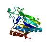
| ||||||||
|---|---|---|---|---|---|---|---|---|---|
| 1 |
| ||||||||
| Unit cell |
|
-Components
| #1: Protein | Mass: 20222.242 Da / Num. of mol.: 1 Source method: isolated from a genetically manipulated source Source: (gene. exp.) Pyrococcus horikoshii (archaea) / Plasmid: PET21a / Production host:  |
|---|---|
| #2: Water | ChemComp-HOH /  Mass: 18.015 Da / Num. of mol.: 88 / Source method: isolated from a natural source / Formula: H2O Mass: 18.015 Da / Num. of mol.: 88 / Source method: isolated from a natural source / Formula: H2O |
-Experimental details
-Experiment
| Experiment | Method: X-RAY DIFFRACTION / Number of used crystals: 1 |
|---|
- Sample preparation
| Crystal | Density Matthews: 1.94 Å3/Da / Density % sol: 36.46 % | ||||||||||||||||||||||||||||||||||||||||||
|---|---|---|---|---|---|---|---|---|---|---|---|---|---|---|---|---|---|---|---|---|---|---|---|---|---|---|---|---|---|---|---|---|---|---|---|---|---|---|---|---|---|---|---|
| Crystal grow | Temperature: 298 K / Method: vapor diffusion, hanging drop / pH: 4.6 Details: 22.5% PEG3350, 250 mM ammonium acetate, 100 mM sodium acetate, pH 4.6. Seeding., VAPOR DIFFUSION, HANGING DROP, temperature 298K | ||||||||||||||||||||||||||||||||||||||||||
| Crystal grow | *PLUS pH: 7.5 | ||||||||||||||||||||||||||||||||||||||||||
| Components of the solutions | *PLUS
|
-Data collection
| Diffraction | Mean temperature: 100 K |
|---|---|
| Diffraction source | Source: SYNCHROTRON / Site: SSRL  / Beamline: BL7-1 / Wavelength: 1.08 Å / Beamline: BL7-1 / Wavelength: 1.08 Å |
| Detector | Type: MARRESEARCH / Detector: IMAGE PLATE / Date: Mar 1, 2000 |
| Radiation | Monochromator: GRAPHITE / Protocol: SINGLE WAVELENGTH / Monochromatic (M) / Laue (L): M / Scattering type: x-ray |
| Radiation wavelength | Wavelength: 1.08 Å / Relative weight: 1 |
| Reflection | Resolution: 2.05→20 Å / Num. all: 9941 / Num. obs: 9862 / % possible obs: 99.2 % / Observed criterion σ(F): 0 / Observed criterion σ(I): 0 / Redundancy: 4.246 % / Biso Wilson estimate: 6.2 Å2 / Rmerge(I) obs: 0.029 / Net I/σ(I): 42.5 |
| Reflection shell | Resolution: 2.05→2.11 Å / Redundancy: 3.1 % / Rmerge(I) obs: 0.118 / Mean I/σ(I) obs: 17.4 / % possible all: 88 |
| Reflection | *PLUS Rmerge(I) obs: 0.027 |
| Reflection shell | *PLUS % possible obs: 88 % / Rmerge(I) obs: 0.093 |
- Processing
| Software |
| ||||||||||||||||||||
|---|---|---|---|---|---|---|---|---|---|---|---|---|---|---|---|---|---|---|---|---|---|
| Refinement | Method to determine structure: MOLECULAR REPLACEMENT Starting model: a structure of the same protein obtained on MAD data Resolution: 2.05→20 Å / Rfactor Rfree error: 0.008 / Data cutoff high absF: 899942.18 / Data cutoff low absF: 0 / Isotropic thermal model: RESTRAINED / Cross valid method: THROUGHOUT / σ(F): 0 / σ(I): 0 / Stereochemistry target values: Engh $ Huber
| ||||||||||||||||||||
| Displacement parameters | Biso mean: 23.6 Å2
| ||||||||||||||||||||
| Refine analyze |
| ||||||||||||||||||||
| Refinement step | Cycle: LAST / Resolution: 2.05→20 Å
| ||||||||||||||||||||
| LS refinement shell | Resolution: 2.05→2.18 Å / Rfactor Rfree error: 0.031 / Total num. of bins used: 6
| ||||||||||||||||||||
| Xplor file |
| ||||||||||||||||||||
| Software | *PLUS Name: SHELXL-97 / Classification: refinement | ||||||||||||||||||||
| Refinement | *PLUS Num. reflection obs: 9761 / σ(F): 0 / Num. reflection Rfree: 955 / Rfactor obs: 0.181 | ||||||||||||||||||||
| Solvent computation | *PLUS | ||||||||||||||||||||
| Displacement parameters | *PLUS | ||||||||||||||||||||
| Refine LS restraints | *PLUS
|
