 Movie
Movie Controller

[English] 日本語
 Yorodumi
- PDB-6ddp: Mycobacterium tuberculosis Dihydrofolate Reductase complexed with... -
+ Open data

- Basic information
| Entry | Database: PDB / ID: 6ddp | ||||||
|---|---|---|---|---|---|---|---|
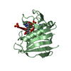
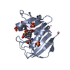
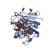
| Title | Mycobacterium tuberculosis Dihydrofolate Reductase complexed with beta-NADPH and 3'-[(2R)-4-(2,4-diamino-6-ethylpyrimidin-5-yl)but-3-yn-2-yl]-5'-methoxy[1,1'-biphenyl]-4-carboxylic acid | ||||||
 Components Components | Dihydrofolate reductase | ||||||
 Keywords Keywords | oxidoreductase/oxidoreductase inhibitor / DHFR / antifolate / oxidoreductase-oxidoreductase inhibitor complex | ||||||
| Function / homology |  Function and homology information Function and homology informationNADP+ binding / dihydrofolate metabolic process / dihydrofolate reductase / dihydrofolate reductase activity / folic acid metabolic process / tetrahydrofolate biosynthetic process / one-carbon metabolic process / NADP binding / cytosol Similarity search - Function | ||||||
| Biological species |   Mycobacterium tuberculosis (bacteria) Mycobacterium tuberculosis (bacteria) | ||||||
| Method |  X-RAY DIFFRACTION / SYNCHROTRON / MOLECULAR REPLACEMENT / Resolution: 1.49 Å X-RAY DIFFRACTION / SYNCHROTRON / MOLECULAR REPLACEMENT / Resolution: 1.49 Å | ||||||
 Authors Authors | Hajian, B. / Wright, D. | ||||||
| Funding support |  United States, 1items United States, 1items
| ||||||
 Citation Citation | Journal: Cell Chem Biol / Year: 2019 Title: Drugging the Folate Pathway in Mycobacterium tuberculosis: The Role of Multi-targeting Agents. Authors: Hajian, B. / Scocchera, E. / Shoen, C. / Krucinska, J. / Viswanathan, K. / G-Dayanandan, N. / Erlandsen, H. / Estrada, A. / Mikusova, K. / Kordulakova, J. / Cynamon, M. / Wright, D. | ||||||
| History |
|
- Structure visualization
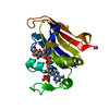
| Structure viewer | Molecule: MolmilJmol/JSmol |
|---|
- Downloads & links
-Download
| PDBx/mmCIF format | 6ddp.cif.gz | 151.4 KB | Display | PDBx/mmCIF format |
|---|---|---|---|---|
| PDB format | pdb6ddp.ent.gz | 119.2 KB | Display | PDB format |
| PDBx/mmJSON format | 6ddp.json.gz | Tree view | PDBx/mmJSON format | |
| Others |  Other downloads Other downloads |
-Validation report
| Arichive directory | https://data.pdbj.org/pub/pdb/validation_reports/dd/6ddpftp://data.pdbj.org/pub/pdb/validation_reports/dd/6ddp | HTTPS FTP |
|---|
-Related structure data
| Related structure data |  6ddsC  6ddwC  6de4C 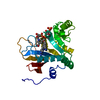 6de5C 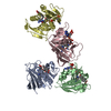 5ja3S S: Starting model for refinement C: citing same article ( |
|---|---|
| Similar structure data |

-Links
 PDBj
PDBj

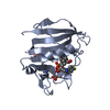
- Assembly
| Deposited unit | 
| ||||||||
|---|---|---|---|---|---|---|---|---|---|
| 1 | 
| ||||||||
| 2 | 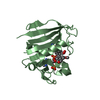
| ||||||||
| 3 | 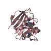
| ||||||||
| 4 | 
| ||||||||
| Unit cell |
|
-Components
| #1: Protein | Mass: 17660.992 Da / Num. of mol.: 4 Source method: isolated from a genetically manipulated source Source: (gene. exp.) Mycobacterium tuberculosis (strain ATCC 25618 / H37Rv) (bacteria)Strain: ATCC 25618 / H37Rv / Gene: folA, dfrA, Rv2763c, MTV002.28c / Production host: #2: Chemical | ChemComp-NDP /   Mass: 745.421 Da / Num. of mol.: 4 / Source method: obtained synthetically / Formula: C21H30N7O17P3 Mass: 745.421 Da / Num. of mol.: 4 / Source method: obtained synthetically / Formula: C21H30N7O17P3#3: Chemical | ChemComp-G6Y / 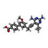  Mass: 416.472 Da / Num. of mol.: 4 / Source method: obtained synthetically / Formula: C24H24N4O3 Mass: 416.472 Da / Num. of mol.: 4 / Source method: obtained synthetically / Formula: C24H24N4O3#4: Water | ChemComp-HOH / |  Mass: 18.015 Da / Num. of mol.: 307 / Source method: isolated from a natural source / Formula: H2O Mass: 18.015 Da / Num. of mol.: 307 / Source method: isolated from a natural source / Formula: H2O |
|---|
-Experimental details
-Experiment
| Experiment | Method: X-RAY DIFFRACTION / Number of used crystals: 1 |
|---|
- Sample preparation
| Crystal | Density Matthews: 3 Å3/Da / Density % sol: 59.04 % |
|---|---|
| Crystal grow | Temperature: 277 K / Method: vapor diffusion Details: 2.1-2.3 M ammonium sulfate, 0.1 M sodium acetate pH 4.5 |
-Data collection
| Diffraction | Mean temperature: 100 K |
|---|---|
| Diffraction source | Source: SYNCHROTRON / Site: SSRL / Beamline: BL14-1 / Wavelength: 0.979 Å |
| Detector | Type: DECTRIS EIGER X 16M / Detector: PIXEL / Date: Jun 19, 2017 |
| Radiation | Protocol: SINGLE WAVELENGTH / Monochromatic (M) / Laue (L): M / Scattering type: x-ray |
| Radiation wavelength | Wavelength: 0.979 Å / Relative weight: 1 |
| Reflection | Resolution: 1.49→34.43 Å / Num. obs: 118402 / % possible obs: 83.15 % / Redundancy: 1.5 % / Rpim(I) all: 0.02 / Rsym value: 0.02 / Net I/σ(I): 19.6 |
| Reflection shell | Resolution: 1.46→1.49 Å / Rpim(I) all: 0.239 / Rsym value: 0.239 |
- Processing
| Software |
| ||||||||||||||||||||||||||||||||||||||||||||||||||||||||||||||||||||||||||||||||||||||||||||||||||||||||||||||||
|---|---|---|---|---|---|---|---|---|---|---|---|---|---|---|---|---|---|---|---|---|---|---|---|---|---|---|---|---|---|---|---|---|---|---|---|---|---|---|---|---|---|---|---|---|---|---|---|---|---|---|---|---|---|---|---|---|---|---|---|---|---|---|---|---|---|---|---|---|---|---|---|---|---|---|---|---|---|---|---|---|---|---|---|---|---|---|---|---|---|---|---|---|---|---|---|---|---|---|---|---|---|---|---|---|---|---|---|---|---|---|---|---|---|
| Refinement | Method to determine structure: MOLECULAR REPLACEMENT Starting model: 5JA3 Resolution: 1.49→34.424 Å / SU ML: 0.16 / Cross valid method: FREE R-VALUE / σ(F): 1.96 / Phase error: 24.61
| ||||||||||||||||||||||||||||||||||||||||||||||||||||||||||||||||||||||||||||||||||||||||||||||||||||||||||||||||
| Solvent computation | Shrinkage radii: 0.9 Å / VDW probe radii: 1.11 Å | ||||||||||||||||||||||||||||||||||||||||||||||||||||||||||||||||||||||||||||||||||||||||||||||||||||||||||||||||
| Refinement step | Cycle: LAST / Resolution: 1.49→34.424 Å
| ||||||||||||||||||||||||||||||||||||||||||||||||||||||||||||||||||||||||||||||||||||||||||||||||||||||||||||||||
| Refine LS restraints |
| ||||||||||||||||||||||||||||||||||||||||||||||||||||||||||||||||||||||||||||||||||||||||||||||||||||||||||||||||
| LS refinement shell |
|
