 Movie
Movie Controller

[English] 日本語
 Yorodumi
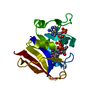
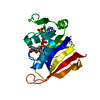
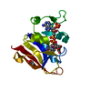
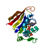
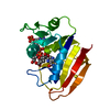
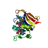
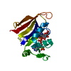
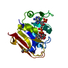
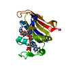
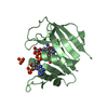
- PDB-3fqv: Staphylococcus aureus F98Y mutant dihydrofolate reductase complex... -
+ Open data

- Basic information
| Entry | Database: PDB / ID: 3fqv | ||||||
|---|---|---|---|---|---|---|---|
| Title | Staphylococcus aureus F98Y mutant dihydrofolate reductase complexed with NADPH and 2,4-diamino-5-[3-(3-methoxy-4-phenylphenyl)but-1-ynyl]-6-methylpyrimidine | ||||||
 Components Components | Trimethoprim-sensitive dihydrofolate reductase | ||||||
 Keywords Keywords | OXIDOREDUCTASE | ||||||
| Function / homology | Dihydrofolate Reductase, subunit A / Dihydrofolate Reductase, subunit A / 3-Layer(aba) Sandwich / Alpha Beta / Chem-11F / Chem-NDP / :  Function and homology information Function and homology information | ||||||
| Biological species |  Staphylococcus aureus RF122 (bacteria) Staphylococcus aureus RF122 (bacteria) | ||||||
| Method |  X-RAY DIFFRACTION / SYNCHROTRON / DIFFERENCE FOURIER / Resolution: 1.85 Å X-RAY DIFFRACTION / SYNCHROTRON / DIFFERENCE FOURIER / Resolution: 1.85 Å | ||||||
 Authors Authors | Anderson, A.C. / Frey, K.M. / Liu, J. / Lombardo, M.N. | ||||||
 Citation Citation | Journal: J.Mol.Biol. / Year: 2009 Title: Crystal structures of wild-type and mutant methicillin-resistant Staphylococcus aureus dihydrofolate reductase reveal an alternate conformation of NADPH that may be linked to trimethoprim resistance. Authors: Frey, K.M. / Liu, J. / Lombardo, M.N. / Bolstad, D.B. / Wright, D.L. / Anderson, A.C. | ||||||
| History |
|
- Structure visualization
| Structure viewer | Molecule: MolmilJmol/JSmol |
|---|
- Downloads & links
-Download
| PDBx/mmCIF format | 3fqv.cif.gz | 53.1 KB | Display | PDBx/mmCIF format |
|---|---|---|---|---|
| PDB format | pdb3fqv.ent.gz | 38.5 KB | Display | PDB format |
| PDBx/mmJSON format | 3fqv.json.gz | Tree view | PDBx/mmJSON format | |
| Others |  Other downloads Other downloads |
-Validation report
| Arichive directory | https://data.pdbj.org/pub/pdb/validation_reports/fq/3fqvftp://data.pdbj.org/pub/pdb/validation_reports/fq/3fqv | HTTPS FTP |
|---|
-Related structure data
| Related structure data | 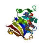 3f0bC  3f0uC 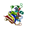 3fq0C  3fqcC  3fqfC 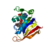 3fqoC  3fqzC C: citing same article ( |
|---|---|
| Similar structure data |


-Links
 PDBj
PDBj

- Assembly
| Deposited unit | 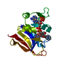
| ||||||||
|---|---|---|---|---|---|---|---|---|---|
| 1 |
| ||||||||
| Unit cell |
|
-Components
| #1: Protein | Mass: 18031.557 Da / Num. of mol.: 1 / Mutation: F98Y Source method: isolated from a genetically manipulated source Source: (gene. exp.) Staphylococcus aureus RF122 (bacteria) / Strain: RF122 / ET3-1 / Gene: dfrB, SAB1281c / Plasmid: pET41 / Production host: |
|---|---|
| #2: Chemical | ChemComp-NDP /   Mass: 745.421 Da / Num. of mol.: 1 / Source method: obtained synthetically / Formula: C21H30N7O17P3 Mass: 745.421 Da / Num. of mol.: 1 / Source method: obtained synthetically / Formula: C21H30N7O17P3 |
| #3: Chemical | ChemComp-11F / 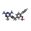  Mass: 358.436 Da / Num. of mol.: 1 / Source method: obtained synthetically / Formula: C22H22N4O Mass: 358.436 Da / Num. of mol.: 1 / Source method: obtained synthetically / Formula: C22H22N4O |
| #4: Water | ChemComp-HOH /  Mass: 18.015 Da / Num. of mol.: 147 / Source method: isolated from a natural source / Formula: H2O Mass: 18.015 Da / Num. of mol.: 147 / Source method: isolated from a natural source / Formula: H2O |
-Experimental details
-Experiment
| Experiment | Method: X-RAY DIFFRACTION / Number of used crystals: 1 |
|---|
- Sample preparation
| Crystal | Density Matthews: 2.7 Å3/Da / Density % sol: 54.37 % |
|---|---|
| Crystal grow | Temperature: 277 K / Method: vapor diffusion, hanging drop / pH: 6.5 Details: 15% PEG 10000, 150mM Sodium acetate, 100mM MES pH 6.5, 5% Butyrlactone, VAPOR DIFFUSION, HANGING DROP, temperature 277K |
-Data collection
| Diffraction | Mean temperature: 77.2 K |
|---|---|
| Diffraction source | Source: SYNCHROTRON / Site: NSLS  / Beamline: X29A / Wavelength: 1.0809 Å / Beamline: X29A / Wavelength: 1.0809 Å |
| Detector | Type: ADSC QUANTUM 315 / Detector: CCD / Date: Jun 20, 2008 |
| Radiation | Protocol: SINGLE WAVELENGTH / Monochromatic (M) / Laue (L): M / Scattering type: x-ray |
| Radiation wavelength | Wavelength: 1.0809 Å / Relative weight: 1 |
| Reflection | Resolution: 1.85→37.06 Å / Num. obs: 16597 / % possible obs: 99.2 % / Observed criterion σ(I): 3 / Redundancy: 10 % / Rmerge(I) obs: 0.047 / Rsym value: 0.047 / Net I/σ(I): 2.7 |
| Reflection shell | Resolution: 1.85→1.92 Å / Redundancy: 8.1 % / Rmerge(I) obs: 0.096 / Mean I/σ(I) obs: 0.9 / Num. unique all: 1203 / Rsym value: 0.096 / % possible all: 99.5 |
- Processing
| Software |
| ||||||||||||||||||||||||||||||||||||||||||||||||||||||||||||||||||||||||||||||||||||||||||
|---|---|---|---|---|---|---|---|---|---|---|---|---|---|---|---|---|---|---|---|---|---|---|---|---|---|---|---|---|---|---|---|---|---|---|---|---|---|---|---|---|---|---|---|---|---|---|---|---|---|---|---|---|---|---|---|---|---|---|---|---|---|---|---|---|---|---|---|---|---|---|---|---|---|---|---|---|---|---|---|---|---|---|---|---|---|---|---|---|---|---|---|
| Refinement | Method to determine structure: DIFFERENCE FOURIER Starting model: Sa F98Y DHFR bound to Folate and NADPH (Dale et al., J.Mol.Biol. 1997, structure not deposited in the PDB) Resolution: 1.85→37.06 Å / Cor.coef. Fo:Fc: 0.927 / Cor.coef. Fo:Fc free: 0.882 / Occupancy max: 1 / Occupancy min: 0.3 / SU B: 3.116 / SU ML: 0.097 / Cross valid method: THROUGHOUT / σ(F): 0 / ESU R: 0.165 / ESU R Free: 0.155 / Stereochemistry target values: MAXIMUM LIKELIHOOD / Details: HYDROGENS HAVE BEEN ADDED IN THE RIDING POSITIONS
| ||||||||||||||||||||||||||||||||||||||||||||||||||||||||||||||||||||||||||||||||||||||||||
| Solvent computation | Ion probe radii: 0.8 Å / Shrinkage radii: 0.8 Å / VDW probe radii: 1.2 Å / Solvent model: MASK | ||||||||||||||||||||||||||||||||||||||||||||||||||||||||||||||||||||||||||||||||||||||||||
| Displacement parameters | Biso max: 60.3 Å2 / Biso mean: 18.144 Å2 / Biso min: 2 Å2
| ||||||||||||||||||||||||||||||||||||||||||||||||||||||||||||||||||||||||||||||||||||||||||
| Refinement step | Cycle: LAST / Resolution: 1.85→37.06 Å
| ||||||||||||||||||||||||||||||||||||||||||||||||||||||||||||||||||||||||||||||||||||||||||
| Refine LS restraints |
| ||||||||||||||||||||||||||||||||||||||||||||||||||||||||||||||||||||||||||||||||||||||||||
| LS refinement shell | Resolution: 1.85→1.92 Å / Total num. of bins used: 20
|
