Protocol: SINGLE WAVELENGTH / Monochromatic (M) / Laue (L): M / Scattering type: x-ray
Radiation wavelength
Wavelength: 0.71 Å / Relative weight: 1
Reflection
Resolution: 1.8→50 Å / Num. obs: 14675 / % possible obs: 95.1 % / Redundancy: 2.3 % / Net I/σ(I): 16.8
-
Processing
Software
Name
Version
Classification
REFMAC
5.7.0032
refinement
HKL-2000
datascaling
Refinement
Resolution: 1.8→44.97 Å / Cor.coef. Fo:Fc: 0.964 / Cor.coef. Fo:Fc free: 0.937 / SU B: 3.571 / SU ML: 0.108 / Cross valid method: THROUGHOUT / ESU R: 0.145 / ESU R Free: 0.151 / Stereochemistry target values: MAXIMUM LIKELIHOOD / Details: HYDROGENS HAVE BEEN ADDED IN THE RIDING POSITIONS
Rfactor
Num. reflection
% reflection
Selection details
Rfree
0.24049
695
5.1 %
RANDOM
Rwork
0.17065
-
-
-
obs
0.17408
13017
95.16 %
-
Solvent computation
Ion probe radii: 0.8 Å / Shrinkage radii: 0.8 Å / VDW probe radii: 1.2 Å / Solvent model: MASK
 Movie
Movie Controller


 Open data
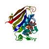
 Basic information
 Components
 Keywords
 Function and homology information
 X-RAY DIFFRACTION /
  Authors
 Citation
 Structure visualization
 Downloads & links
 Other downloads


 PDBj
PDBj


 Assembly
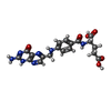
 Mass: 441.397 Da / Num. of mol.: 1 / Source method: obtained synthetically / Formula: C19H19N7O6
 Mass: 743.405 Da / Num. of mol.: 1 / Source method: obtained synthetically / Formula: C21H28N7O17P3
 Mass: 78.133 Da / Num. of mol.: 1 / Source method: obtained synthetically / Formula: C2H6OS
 Mass: 18.015 Da / Num. of mol.: 137 / Source method: isolated from a natural source / Formula: H2O
 Sample preparation
 / Beamline: AR-NW12A / Wavelength: 0.71 Å
 Processing