 Movie
Movie Controller

[English] 日本語
 Yorodumi
- PDB-1l7z: Crystal structure of Ca2+/Calmodulin complexed with myristoylated... -
+ Open data

- Basic information
| Entry | Database: PDB / ID: 1l7z | ||||||
|---|---|---|---|---|---|---|---|
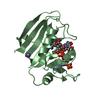
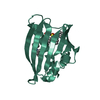
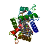
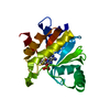
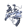
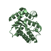
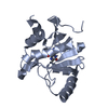
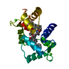
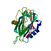
| Title | Crystal structure of Ca2+/Calmodulin complexed with myristoylated CAP-23/NAP-22 peptide | ||||||
 Components Components |
| ||||||
 Keywords Keywords | METAL BINDING PROTEIN/PROTEIN BINDING / CALMODULIN / MYRISTOYLATION / PROTEIN-PROTEIN INTERACTION / RIKEN Structural Genomics/Proteomics Initiative / RSGI / Structural Genomics / METAL BINDING PROTEIN-PROTEIN BINDING COMPLEX | ||||||
| Function / homology |  Function and homology information Function and homology information: / : / : / : / positive regulation of protein autophosphorylation / : / negative regulation of peptidyl-threonine phosphorylation / : / type 3 metabotropic glutamate receptor binding / positive regulation of peptidyl-threonine phosphorylation ...: / : / : / : / positive regulation of protein autophosphorylation / : / negative regulation of peptidyl-threonine phosphorylation / : / type 3 metabotropic glutamate receptor binding / positive regulation of peptidyl-threonine phosphorylation / positive regulation of DNA binding / CaM pathway / Cam-PDE 1 activation / Sodium/Calcium exchangers / Calmodulin induced events / Reduction of cytosolic Ca++ levels / Activation of Ca-permeable Kainate Receptor / CREB1 phosphorylation through the activation of CaMKII/CaMKK/CaMKIV cascasde / Loss of phosphorylation of MECP2 at T308 / CREB1 phosphorylation through the activation of Adenylate Cyclase / negative regulation of high voltage-gated calcium channel activity / PKA activation / CaMK IV-mediated phosphorylation of CREB / Glycogen breakdown (glycogenolysis) / CLEC7A (Dectin-1) induces NFAT activation / response to corticosterone / negative regulation of ryanodine-sensitive calcium-release channel activity / organelle localization by membrane tethering / Activation of RAC1 downstream of NMDARs / : / autophagosome membrane docking / regulation of synaptic vesicle exocytosis / negative regulation of calcium ion export across plasma membrane / regulation of ryanodine-sensitive calcium-release channel activity / regulation of cardiac muscle cell action potential / presynaptic endocytosis / Synthesis of IP3 and IP4 in the cytosol / positive regulation of protein serine/threonine kinase activity / Phase 0 - rapid depolarisation / Negative regulation of NMDA receptor-mediated neuronal transmission / Unblocking of NMDA receptors, glutamate binding and activation / RHO GTPases activate PAKs / calcineurin-mediated signaling / nitric-oxide synthase binding / regulation of cell communication by electrical coupling involved in cardiac conduction / Ion transport by P-type ATPases / adenylate cyclase binding / Uptake and function of anthrax toxins / protein phosphatase activator activity / Long-term potentiation / Calcineurin activates NFAT / Regulation of MECP2 expression and activity / DARPP-32 events / Smooth Muscle Contraction / regulation of synaptic vesicle endocytosis / detection of calcium ion / regulation of cardiac muscle contraction / catalytic complex / RHO GTPases activate IQGAPs / phosphatidylinositol 3-kinase binding / positive regulation of nitric-oxide synthase activity / activation of adenylate cyclase activity / calcium channel inhibitor activity / presynaptic cytosol / Activation of AMPK downstream of NMDARs / cellular response to interferon-beta / enzyme regulator activity / regulation of release of sequestered calcium ion into cytosol by sarcoplasmic reticulum / Ion homeostasis / eNOS activation / Tetrahydrobiopterin (BH4) synthesis, recycling, salvage and regulation / titin binding / Protein methylation / regulation of cardiac muscle contraction by regulation of the release of sequestered calcium ion / regulation of calcium-mediated signaling / voltage-gated potassium channel complex / FCERI mediated Ca+2 mobilization / calcium channel complex / substantia nigra development / FCGR3A-mediated IL10 synthesis / regulation of heart rate / Antigen activates B Cell Receptor (BCR) leading to generation of second messengers / calyx of Held / Ras activation upon Ca2+ influx through NMDA receptor / nitric-oxide synthase regulator activity / adenylate cyclase activator activity / VEGFR2 mediated cell proliferation / VEGFR2 mediated vascular permeability / regulation of cytokinesis / protein serine/threonine kinase activator activity / spindle microtubule / positive regulation of receptor signaling pathway via JAK-STAT / sarcomere / response to amphetamine / Translocation of SLC2A4 (GLUT4) to the plasma membrane / calcium channel regulator activity / Transcriptional activation of mitochondrial biogenesis / RAF activation / response to calcium ion / cellular response to type II interferon Similarity search - Function | ||||||
| Biological species |  Homo sapiens (human) Homo sapiens (human) | ||||||
| Method |  X-RAY DIFFRACTION / SYNCHROTRON / MOLECULAR REPLACEMENT / Resolution: 2.3 Å X-RAY DIFFRACTION / SYNCHROTRON / MOLECULAR REPLACEMENT / Resolution: 2.3 Å | ||||||
 Authors Authors | Matsubara, M. / Nakatsu, T. / Yamauchi, E. / Kato, H. / Taniguchi, H. / RIKEN Structural Genomics/Proteomics Initiative (RSGI) | ||||||
 Citation Citation | Journal: EMBO J. / Year: 2004 Title: Crystal structure of a myristoylated CAP-23/NAP-22 N-terminal domain complexed with Ca2+/calmodulin Authors: Matsubara, M. / Nakatsu, T. / Kato, H. / Taniguchi, H. #1: Journal: J.Biol.Chem. / Year: 1999 Title: Identification of the calmodulin-binding domain of neuron-specific protein kinase C substrate protein CAP-22/NAP-22. Direct involvement of protein myristoylation in calmodulin-target protein interaction Authors: Takasaki, A. / Hayashi, N. / Matsubara, M. / Yamauchi, E. / Taniguchi, H. | ||||||
| History |
|
- Structure visualization
| Structure viewer | Molecule: MolmilJmol/JSmol |
|---|
- Downloads & links
-Download
| PDBx/mmCIF format | 1l7z.cif.gz | 46.5 KB | Display | PDBx/mmCIF format |
|---|---|---|---|---|
| PDB format | pdb1l7z.ent.gz | 31.8 KB | Display | PDB format |
| PDBx/mmJSON format | 1l7z.json.gz | Tree view | PDBx/mmJSON format | |
| Others |  Other downloads Other downloads |
-Validation report
| Arichive directory | https://data.pdbj.org/pub/pdb/validation_reports/l7/1l7zftp://data.pdbj.org/pub/pdb/validation_reports/l7/1l7z | HTTPS FTP |
|---|
-Related structure data
| Related structure data |  1qivS S: Starting model for refinement |
|---|---|
| Similar structure data | |
| Other databases |

-Links
 PDBj
PDBj


- Assembly
| Deposited unit | 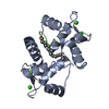
| ||||||||
|---|---|---|---|---|---|---|---|---|---|
| 1 |
| ||||||||
| Unit cell |
|
-Components
| #1: Protein | Mass: 16721.350 Da / Num. of mol.: 1 Source method: isolated from a genetically manipulated source Source: (gene. exp.) Homo sapiens (human) / Plasmid: pACYC/hCaM / Species (production host): Escherichia coli / Production host:  | ||||||
|---|---|---|---|---|---|---|---|
| #2: Protein/peptide | Mass: 978.253 Da / Num. of mol.: 1 / Fragment: calmodulin binding domain / Source method: obtained synthetically / Details: synthetic myristoylated peptide / References: GenBank: 220820 | ||||||
| #3: Chemical | ChemComp-CA /   Mass: 40.078 Da / Num. of mol.: 4 / Source method: obtained synthetically / Formula: Ca Mass: 40.078 Da / Num. of mol.: 4 / Source method: obtained synthetically / Formula: Ca#4: Chemical | ChemComp-MYR / |   Mass: 228.371 Da / Num. of mol.: 1 / Source method: obtained synthetically / Formula: C14H28O2 Mass: 228.371 Da / Num. of mol.: 1 / Source method: obtained synthetically / Formula: C14H28O2#5: Water | ChemComp-HOH / |  Mass: 18.015 Da / Num. of mol.: 75 / Source method: isolated from a natural source / Formula: H2O Mass: 18.015 Da / Num. of mol.: 75 / Source method: isolated from a natural source / Formula: H2OHas protein modification | Y | |
-Experimental details
-Experiment
| Experiment | Method: X-RAY DIFFRACTION / Number of used crystals: 1 |
|---|
- Sample preparation
| Crystal | Density Matthews: 2.33 Å3/Da / Density % sol: 47.3 % | ||||||||||||||||||||||||||||||||||||||||||
|---|---|---|---|---|---|---|---|---|---|---|---|---|---|---|---|---|---|---|---|---|---|---|---|---|---|---|---|---|---|---|---|---|---|---|---|---|---|---|---|---|---|---|---|
| Crystal grow | Temperature: 277 K / Method: vapor diffusion, hanging drop / pH: 5.5 Details: PEG 1000, cacodylate, pH 5.5, VAPOR DIFFUSION, HANGING DROP, temperature 277K | ||||||||||||||||||||||||||||||||||||||||||
| Crystal grow | *PLUS Temperature: 4 ℃ / Method: vapor diffusion, hanging drop | ||||||||||||||||||||||||||||||||||||||||||
| Components of the solutions | *PLUS
|
-Data collection
| Diffraction | Mean temperature: 90 K |
|---|---|
| Diffraction source | Source: SYNCHROTRON / Site: SPring-8  / Beamline: BL45PX / Wavelength: 1.02 Å / Beamline: BL45PX / Wavelength: 1.02 Å |
| Detector | Type: RIGAKU RAXIS V / Detector: IMAGE PLATE / Date: Nov 7, 2000 |
| Radiation | Monochromator: DIAMOND / Protocol: SINGLE WAVELENGTH / Monochromatic (M) / Laue (L): M / Scattering type: x-ray |
| Radiation wavelength | Wavelength: 1.02 Å / Relative weight: 1 |
| Reflection | Resolution: 2.3→50 Å / Num. all: 7815 / Num. obs: 7715 / % possible obs: 96.9 % / Observed criterion σ(F): 1 / Observed criterion σ(I): 1 / Redundancy: 5.1 % / Biso Wilson estimate: 51.5 Å2 / Rsym value: 0.083 / Net I/σ(I): 14.7 |
| Reflection shell | Resolution: 2.3→2.38 Å / Num. unique all: 793 / Rsym value: 0.192 / % possible all: 99.2 |
| Reflection | *PLUS Num. obs: 7815 / Num. measured all: 39942 / Rmerge(I) obs: 0.083 |
- Processing
| Software |
| |||||||||||||||||||||||||
|---|---|---|---|---|---|---|---|---|---|---|---|---|---|---|---|---|---|---|---|---|---|---|---|---|---|---|
| Refinement | Method to determine structure: MOLECULAR REPLACEMENT Starting model: PDB ENTRY 1QIV Resolution: 2.3→20 Å / σ(F): 2 / Stereochemistry target values: Engh & Huber
| |||||||||||||||||||||||||
| Refine analyze |
| |||||||||||||||||||||||||
| Refinement step | Cycle: LAST / Resolution: 2.3→20 Å
| |||||||||||||||||||||||||
| Refine LS restraints |
| |||||||||||||||||||||||||
| Refinement | *PLUS Lowest resolution: 20 Å / Num. reflection obs: 7799 / % reflection Rfree: 5 % / Rfactor Rfree: 0.274 / Rfactor Rwork: 0.247 | |||||||||||||||||||||||||
| Solvent computation | *PLUS | |||||||||||||||||||||||||
| Displacement parameters | *PLUS | |||||||||||||||||||||||||
| Refine LS restraints | *PLUS
|
