 Movie
Movie Controller


+ Open data

- Basic information
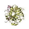
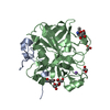
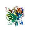
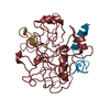
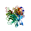
| Entry | Database: PDB / ID: 3edx | ||||||
|---|---|---|---|---|---|---|---|
| Title | Crystal structure of the W215A/E217A mutant of murine thrombin | ||||||
 Components Components |
| ||||||
 Keywords Keywords |  HYDROLASE / Serine protease / Acute phase / Blood coagulation / Cleavage on pair of basic residues / Gamma-carboxyglutamic acid / Glycoprotein / Kringle / Protease / Zymogen HYDROLASE / Serine protease / Acute phase / Blood coagulation / Cleavage on pair of basic residues / Gamma-carboxyglutamic acid / Glycoprotein / Kringle / Protease / Zymogen | ||||||
| Function / homology |  Function and homology information Function and homology informationCommon Pathway of Fibrin Clot Formation / Platelet Aggregation (Plug Formation) / Gamma-carboxylation of protein precursors / Transport of gamma-carboxylated protein precursors from the endoplasmic reticulum to the Golgi apparatus / Intrinsic Pathway of Fibrin Clot Formation / Removal of aminoterminal propeptides from gamma-carboxylated proteins / thrombin-activated receptor signaling pathway / Peptide ligand-binding receptors / Thrombin signalling through proteinase activated receptors (PARs) / Regulation of Complement cascade ...Common Pathway of Fibrin Clot Formation / Platelet Aggregation (Plug Formation) / Gamma-carboxylation of protein precursors / Transport of gamma-carboxylated protein precursors from the endoplasmic reticulum to the Golgi apparatus / Intrinsic Pathway of Fibrin Clot Formation / Removal of aminoterminal propeptides from gamma-carboxylated proteins / thrombin-activated receptor signaling pathway / Peptide ligand-binding receptors / Thrombin signalling through proteinase activated receptors (PARs) / Regulation of Complement cascade / G alpha (q) signalling events / Cell surface interactions at the vascular wall / positive regulation of phospholipase C-activating G protein-coupled receptor signaling pathway / cytolysis by host of symbiont cells / thrombospondin receptor activity / thrombin / neutrophil-mediated killing of gram-negative bacterium / ligand-gated ion channel signaling pathway / negative regulation of astrocyte differentiation / positive regulation of collagen biosynthetic process / negative regulation of cytokine production involved in inflammatory response / positive regulation of blood coagulation / fibrinolysis / regulation of cytosolic calcium ion concentration / positive regulation of release of sequestered calcium ion into cytosol / acute-phase response / lipopolysaccharide binding / negative regulation of proteolysis / positive regulation of insulin secretion / platelet activation / response to wounding / positive regulation of protein localization to nucleus / positive regulation of reactive oxygen species metabolic process / antimicrobial humoral immune response mediated by antimicrobial peptide / heparin binding / peptidase activity / regulation of cell shape / positive regulation of cell growth / regulation of gene expression / collagen-containing extracellular matrix / endopeptidase activity / cell surface receptor signaling pathway / positive regulation of phosphatidylinositol 3-kinase/protein kinase B signal transduction / positive regulation of protein phosphorylation / G protein-coupled receptor signaling pathway / external side of plasma membrane / signaling receptor binding / serine-type endopeptidase activity / calcium ion binding / positive regulation of cell population proliferation / proteolysis / extracellular spaceSimilarity search - Function | ||||||
| Biological species |  Mus musculus (house mouse) Mus musculus (house mouse) | ||||||
| Method | X-RAY DIFFRACTION / SYNCHROTRON / MOLECULAR REPLACEMENT / Resolution: 2.4 Å | ||||||
 Authors Authors | Gandhi, P.S. / Page, M.J. / Chen, Z. / Bush-pelc, L. / Di Cera, E. | ||||||
 Citation Citation | Journal: To be Published Title: Molecular Basis for the Kinetic Differences of the W215A/E217A Mutant of Human and Murine Thrombin Authors: Gandhi, P.S. / Page, M.J. / Chen, Z. / Bush-Pelc, L. / Di Cera, E. #1: Journal: J.Biol.Chem. / Year: 2004Title: The Anticoagulant Thrombin Mutant W215/E217A Has a Collapsed Primary Specificity Pocket Authors: Pineda, A.O. / Chen, Z. / Caccia, S. / Cantwell, A.M. / Savvides, S.N. / Waksman, G. / Mathews, F.S. / Di Cera, E. | ||||||
| History |
|
- Structure visualization
| Structure viewer | Molecule: MolmilJmol/JSmol |
|---|
- Downloads & links
-Download
| PDBx/mmCIF format | 3edx.cif.gz | 198.3 KB | Display | PDBx/mmCIF format |
|---|---|---|---|---|
| PDB format | pdb3edx.ent.gz | 158.8 KB | Display | PDB format |
| PDBx/mmJSON format | 3edx.json.gz | Tree view | PDBx/mmJSON format | |
| Others |  Other downloads Other downloads |
-Validation report
| Arichive directory | https://data.pdbj.org/pub/pdb/validation_reports/ed/3edxftp://data.pdbj.org/pub/pdb/validation_reports/ed/3edx | HTTPS FTP |
|---|
-Related structure data
| Related structure data |  3ee0C  1tq0S S: Starting model for refinement C: citing same article ( |
|---|---|
| Similar structure data |


-Links
 PDBj
PDBj


- Assembly
| Deposited unit | 
| ||||||||
|---|---|---|---|---|---|---|---|---|---|
| 1 | 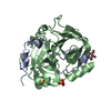
| ||||||||
| 2 | 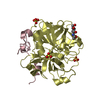
| ||||||||
| 3 | 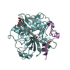
| ||||||||
| Unit cell |
|
-Components
| #1: Protein/peptide | Mass: 5105.731 Da / Num. of mol.: 3 Source method: isolated from a genetically manipulated source Source: (gene. exp.) Mus musculus (house mouse) / Gene: F2, Cf2 / Plasmid: HPC4-pNUT / Production host:  Cricetulus griseus (Chinese hamster) / References: UniProt: P19221, thrombin Cricetulus griseus (Chinese hamster) / References: UniProt: P19221, thrombin#2: Protein | Mass: 29795.461 Da / Num. of mol.: 3 / Mutation: W215A, E217A Source method: isolated from a genetically manipulated source Source: (gene. exp.) Mus musculus (house mouse) / Gene: F2, Cf2 / Plasmid: HPC4-pNUT / Production host: Cricetulus griseus (Chinese hamster) / References: UniProt: P19221, thrombin#3: Chemical | ChemComp-SO4 / Sulfate  Mass: 96.063 Da / Num. of mol.: 7 / Source method: obtained synthetically / Formula: SO4 Mass: 96.063 Da / Num. of mol.: 7 / Source method: obtained synthetically / Formula: SO4#4: Sugar | N-Acetylglucosamine  Type: D-saccharide, beta linking / Mass: 221.208 Da / Num. of mol.: 3 Type: D-saccharide, beta linking / Mass: 221.208 Da / Num. of mol.: 3Source method: isolated from a genetically manipulated source Formula: C8H15NO6 #5: Water | ChemComp-HOH / | Water Mass: 18.015 Da / Num. of mol.: 227 / Source method: isolated from a natural source / Formula: H2O Mass: 18.015 Da / Num. of mol.: 227 / Source method: isolated from a natural source / Formula: H2O |
|---|
-Experimental details
-Experiment
| Experiment | Method: X-RAY DIFFRACTION / Number of used crystals: 1 |
|---|
- Sample preparation
| Crystal | Density Matthews: 4.36 Å3/Da / Density % sol: 71.81 % |
|---|---|
| Crystal grow | Temperature: 295 K / Method: vapor diffusion, hanging drop / pH: 6.6 Details: 0.2M NA2SO4, 20 % PEG 3350, pH 6.6, VAPOR DIFFUSION, HANGING DROP, temperature 295K |
-Data collection
| Diffraction | Mean temperature: 100 K |
|---|---|
| Diffraction source | Source: SYNCHROTRON / Site: APS  / Beamline: 14-BM-C / Wavelength: 0.9 Å / Beamline: 14-BM-C / Wavelength: 0.9 Å |
| Detector | Type: ADSC QUANTUM 315 / Detector: CCD / Date: Apr 14, 2006 |
| Radiation | Protocol: SINGLE WAVELENGTH / Monochromatic (M) / Laue (L): M / Scattering type: x-ray |
| Radiation wavelength | Wavelength: 0.9 Å / Relative weight: 1 |
| Reflection | Resolution: 2.4→40 Å / Num. all: 71558 / Num. obs: 67622 / % possible obs: 94.5 % / Observed criterion σ(F): -1 / Observed criterion σ(I): -1 / Redundancy: 5.6 % / Biso Wilson estimate: 33.3 Å2 / Rmerge(I) obs: 0.059 / Net I/σ(I): 21.2 |
| Reflection shell | Resolution: 2.4→2.49 Å / Redundancy: 4.1 % / Rmerge(I) obs: 0.461 / Mean I/σ(I) obs: 2.5 / Num. unique all: 5642 / % possible all: 79.9 |
- Processing
| Software |
| |||||||||||||||||||||||||
|---|---|---|---|---|---|---|---|---|---|---|---|---|---|---|---|---|---|---|---|---|---|---|---|---|---|---|
| Refinement | Method to determine structure: MOLECULAR REPLACEMENT Starting model: PDB entry 1TQ0 Resolution: 2.4→32.78 Å / Rfactor Rfree error: 0.004 / Data cutoff high absF: 219529.95 / Data cutoff low absF: 0 / Isotropic thermal model: Isotropic / Cross valid method: THROUGHOUT / σ(F): 0 / σ(I): 0 / Stereochemistry target values: Engh & Huber
| |||||||||||||||||||||||||
| Displacement parameters | Biso mean: 55.6 Å2 | |||||||||||||||||||||||||
| Refine analyze |
| |||||||||||||||||||||||||
| Refinement step | Cycle: LAST / Resolution: 2.4→32.78 Å
| |||||||||||||||||||||||||
| Refine LS restraints |
| |||||||||||||||||||||||||
| LS refinement shell | Resolution: 2.4→2.55 Å / Rfactor Rfree error: 0.016 / Total num. of bins used: 6
|
