 Movie
Movie Controller

[English] 日本語
 Yorodumi
- PDB-3eb4: Voltage-dependent K+ channel beta subunit (I211R) in complex with... -
+ Open data

- Basic information
| Entry | Database: PDB / ID: 3eb4 | ||||||
|---|---|---|---|---|---|---|---|
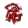
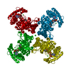
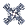
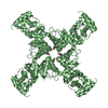
| Title | Voltage-dependent K+ channel beta subunit (I211R) in complex with cortisone | ||||||
 Components Components | Voltage-gated potassium channel subunit beta-2 | ||||||
 Keywords Keywords | TRANSPORT PROTEIN / oxidoreductase / kvbeta / cortisone / interface / Cytoplasm / Ion transport / Ionic channel / NADP / Phosphoprotein / Potassium / Potassium transport / Transport / Voltage-gated channel | ||||||
| Function / homology |  Function and homology information Function and homology informationpinceau fiber / Voltage gated Potassium channels / methylglyoxal reductase (NADPH) (acetol producing) activity / potassium channel complex / regulation of protein localization to cell surface / : / axon initial segment / Oxidoreductases; Acting on the CH-OH group of donors; With NAD+ or NADP+ as acceptor / juxtaparanode region of axon / myoblast differentiation ...pinceau fiber / Voltage gated Potassium channels / methylglyoxal reductase (NADPH) (acetol producing) activity / potassium channel complex / regulation of protein localization to cell surface / : / axon initial segment / Oxidoreductases; Acting on the CH-OH group of donors; With NAD+ or NADP+ as acceptor / juxtaparanode region of axon / myoblast differentiation / regulation of potassium ion transmembrane transport / Neutrophil degranulation / neuromuscular process / voltage-gated potassium channel activity / potassium channel regulator activity / hematopoietic progenitor cell differentiation / voltage-gated potassium channel complex / axon terminus / postsynaptic density membrane / cytoplasmic side of plasma membrane / transmembrane transporter binding / cytoskeleton / neuron projection / postsynaptic density / axon / protein-containing complex binding / glutamatergic synapse / membrane / cytosol Similarity search - Function | ||||||
| Biological species |  | ||||||
| Method |  X-RAY DIFFRACTION / SYNCHROTRON / MOLECULAR REPLACEMENT / Resolution: 2 Å X-RAY DIFFRACTION / SYNCHROTRON / MOLECULAR REPLACEMENT / Resolution: 2 Å | ||||||
 Authors Authors | Pan, Y. / Weng, J. / Kabaleeswaran, V. / Li, H. / Cao, Y. / Bhosle, R.C. / Zhou, M. | ||||||
 Citation Citation | Journal: Nat.Chem.Biol. / Year: 2008 Title: Cortisone dissociates the Shaker family K+ channels from their beta subunits. Authors: Pan, Y. / Weng, J. / Kabaleeswaran, V. / Li, H. / Cao, Y. / Bhosle, R.C. / Zhou, M. #1: Journal: Cell(Cambridge,Mass.) / Year: 1999Title: Structure of a voltage-dependent K+ channel beta subunit. Authors: Gulbis, J.M. / Mann, S. / MacKinnon, R. #2: Journal: Science / Year: 2000Title: Structure of the cytoplasmic beta subunit-T1 assembly of voltage-dependent K+ channels. Authors: Gulbis, J.M. / Zhou, M. / Mann, S. / MacKinnon, R. | ||||||
| History |
|
- Structure visualization
| Structure viewer | Molecule: MolmilJmol/JSmol |
|---|
- Downloads & links
-Download
| PDBx/mmCIF format | 3eb4.cif.gz | 84.1 KB | Display | PDBx/mmCIF format |
|---|---|---|---|---|
| PDB format | pdb3eb4.ent.gz | 61.8 KB | Display | PDB format |
| PDBx/mmJSON format | 3eb4.json.gz | Tree view | PDBx/mmJSON format | |
| Others |  Other downloads Other downloads |
-Validation report
| Arichive directory | https://data.pdbj.org/pub/pdb/validation_reports/eb/3eb4ftp://data.pdbj.org/pub/pdb/validation_reports/eb/3eb4 | HTTPS FTP |
|---|
-Related structure data
| Related structure data | 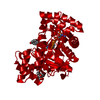 3eauC  3eb3C 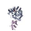 1exbS S: Starting model for refinement C: citing same article ( |
|---|---|
| Similar structure data |


-Links
 PDBj
PDBj


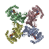
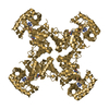
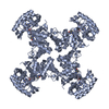
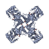
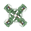
- Assembly
| Deposited unit | 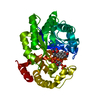
| ||||||||
|---|---|---|---|---|---|---|---|---|---|
| 1 | 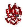
| ||||||||
| Unit cell |
|
-Components
| #1: Protein | Mass: 36616.227 Da / Num. of mol.: 1 / Fragment: cytoplasmic Kvbeta subunit / Mutation: I211R Source method: isolated from a genetically manipulated source Source: (gene. exp.)  |
|---|---|
| #2: Chemical | ChemComp-NDP /   Mass: 745.421 Da / Num. of mol.: 1 / Source method: obtained synthetically / Formula: C21H30N7O17P3 Mass: 745.421 Da / Num. of mol.: 1 / Source method: obtained synthetically / Formula: C21H30N7O17P3 |
| #3: Chemical | ChemComp-PDN /   Mass: 358.428 Da / Num. of mol.: 1 / Source method: obtained synthetically / Formula: C21H26O5 / Comment: medication*YM Mass: 358.428 Da / Num. of mol.: 1 / Source method: obtained synthetically / Formula: C21H26O5 / Comment: medication*YM |
| #4: Water | ChemComp-HOH /  Mass: 18.015 Da / Num. of mol.: 160 / Source method: isolated from a natural source / Formula: H2O Mass: 18.015 Da / Num. of mol.: 160 / Source method: isolated from a natural source / Formula: H2O |
-Experimental details
-Experiment
| Experiment | Method: X-RAY DIFFRACTION / Number of used crystals: 1 |
|---|
- Sample preparation
| Crystal | Density Matthews: 3.22 Å3/Da / Density % sol: 61.85 % |
|---|---|
| Crystal grow | Temperature: 293 K / Method: vapor diffusion, sitting drop Details: 6-15% glycerol, 1.5 M ammonium sulfate, 0.1 M Tris, VAPOR DIFFUSION, SITTING DROP, temperature 293K PH range: 7.9 - 8.8 |
-Data collection
| Diffraction | Mean temperature: 100 K |
|---|---|
| Diffraction source | Source: SYNCHROTRON / Site: NSLS  / Beamline: X4C / Wavelength: 0.97893 Å / Beamline: X4C / Wavelength: 0.97893 Å |
| Detector | Type: MAR scanner 345 mm plate / Detector: IMAGE PLATE / Date: Aug 26, 2007 |
| Radiation | Monochromator: single Si (111) / Protocol: SINGLE WAVELENGTH / Monochromatic (M) / Laue (L): M / Scattering type: x-ray |
| Radiation wavelength | Wavelength: 0.97893 Å / Relative weight: 1 |
| Reflection | Resolution: 1.95→50 Å / Num. obs: 35198 / % possible obs: 100 % / Observed criterion σ(F): 1.5 / Observed criterion σ(I): 2 / Redundancy: 10.6 % / Biso Wilson estimate: 16.3 Å2 / Rmerge(I) obs: 0.064 / Rsym value: 0.045 / Net I/σ(I): 35.66 |
| Reflection shell | Resolution: 1.95→2.02 Å / Redundancy: 9.4 % / Rmerge(I) obs: 0.46 / Mean I/σ(I) obs: 4.48 / Num. unique all: 3063 / % possible all: 100 |
- Processing
| Software |
| ||||||||||||||||||||||||||||||||||||
|---|---|---|---|---|---|---|---|---|---|---|---|---|---|---|---|---|---|---|---|---|---|---|---|---|---|---|---|---|---|---|---|---|---|---|---|---|---|
| Refinement | Method to determine structure: MOLECULAR REPLACEMENT Starting model: pdb entry 1EXB, chain A, beta subunit Resolution: 2→40.04 Å / Rfactor Rfree error: 0.006 / Data cutoff high absF: 3391309.07 / Data cutoff low absF: 0 / Isotropic thermal model: RESTRAINED / Cross valid method: THROUGHOUT / σ(F): 2 / σ(I): 2 / Stereochemistry target values: Engh & Huber
| ||||||||||||||||||||||||||||||||||||
| Solvent computation | Solvent model: FLAT MODEL / Bsol: 44.3035 Å2 / ksol: 0.35606 e/Å3 | ||||||||||||||||||||||||||||||||||||
| Displacement parameters | Biso mean: 29.4 Å2
| ||||||||||||||||||||||||||||||||||||
| Refine analyze |
| ||||||||||||||||||||||||||||||||||||
| Refinement step | Cycle: LAST / Resolution: 2→40.04 Å
| ||||||||||||||||||||||||||||||||||||
| Refine LS restraints |
| ||||||||||||||||||||||||||||||||||||
| LS refinement shell | Resolution: 2→2.07 Å / Rfactor Rfree error: 0.026 / Total num. of bins used: 6
| ||||||||||||||||||||||||||||||||||||
| Xplor file |
|
