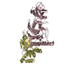
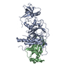
- PDB-3e0j: X-ray structure of the complex of regulatory subunits of human DN... -
+
Open data
ID or keywords:
Loading...
-
Basic information
Entry
Database: PDB / ID: 3e0j
Title
X-ray structure of the complex of regulatory subunits of human DNA polymerase delta
Components
DNA polymerase subunit delta-2
DNA polymerase subunit delta-3
Keywords
TRANSFERASE / DNA Polymerase Delta / p66 subunit / p50 subunit / human / DNA replication / DNA-directed DNA polymerase / Nucleotidyltransferase / Nucleus / Polymorphism / Phosphoprotein
Function / homology
Function and homology information
delta DNA polymerase complex / DNA synthesis involved in UV-damage excision repair / zeta DNA polymerase complex / nucleotide-excision repair, DNA gap filling / Polymerase switching / Processive synthesis on the lagging strand / Removal of the Flap Intermediate / Telomere C-strand (Lagging Strand) Synthesis / Mismatch repair (MMR) directed by MSH2:MSH3 (MutSbeta) / Mismatch repair (MMR) directed by MSH2:MSH6 (MutSalpha) ...delta DNA polymerase complex / DNA synthesis involved in UV-damage excision repair / zeta DNA polymerase complex / nucleotide-excision repair, DNA gap filling / Polymerase switching / Processive synthesis on the lagging strand / Removal of the Flap Intermediate / Telomere C-strand (Lagging Strand) Synthesis / Mismatch repair (MMR) directed by MSH2:MSH3 (MutSbeta) / Mismatch repair (MMR) directed by MSH2:MSH6 (MutSalpha) / Polymerase switching on the C-strand of the telomere / Processive synthesis on the C-strand of the telomere / Removal of the Flap Intermediate from the C-strand / DNA biosynthetic process / DNA strand elongation involved in DNA replication / DNA synthesis involved in DNA repair / PCNA-Dependent Long Patch Base Excision Repair / error-prone translesion synthesis / mismatch repair / Gap-filling DNA repair synthesis and ligation in GG-NER / Termination of translesion DNA synthesis / Recognition of DNA damage by PCNA-containing replication complex / HDR through Homologous Recombination (HRR) / DNA-templated DNA replication / Dual Incision in GG-NER / Dual incision in TC-NER / Gap-filling DNA repair synthesis and ligation in TC-NER / protein-macromolecule adaptor activity / DNA replication / DNA binding / nucleoplasm / nucleus / cytoplasm Similarity search - Function
DNA polymerase delta, p66 (Cdc27) subunit, wHTH domain / OB fold (Dihydrolipoamide Acetyltransferase, E2P) - #430 / Purple Acid Phosphatase; chain A, domain 2 - #50 / DNA polymerase delta subunit 3 / DNA polymerase delta subunit 3 superfamily / DNA polymerase subunit Cdc27 / DNA polymerase delta subunit, OB-fold domain / DNA polymerase delta subunit 2, C-terminal domain / DNA polymerase delta subunit OB-fold domain / DNA polymerase delta/II small subunit family ...DNA polymerase delta, p66 (Cdc27) subunit, wHTH domain / OB fold (Dihydrolipoamide Acetyltransferase, E2P) - #430 / Purple Acid Phosphatase; chain A, domain 2 - #50 / DNA polymerase delta subunit 3 / DNA polymerase delta subunit 3 superfamily / DNA polymerase subunit Cdc27 / DNA polymerase delta subunit, OB-fold domain / DNA polymerase delta subunit 2, C-terminal domain / DNA polymerase delta subunit OB-fold domain / DNA polymerase delta/II small subunit family / 50s Ribosomal Protein L17; Chain: A, / DNA polymerase alpha/delta/epsilon, subunit B / DNA polymerase alpha/epsilon subunit B / Purple Acid Phosphatase; chain A, domain 2 / 4-Layer Sandwich / OB fold (Dihydrolipoamide Acetyltransferase, E2P) / Alpha-Beta Complex / Beta Barrel / Mainly Beta / Alpha Beta Similarity search - Domain/homology
#1: Journal: Acta Crystallogr.,Sect.F / Year: 2008 Title: Crystallization and preliminary crystallographic analysis of the complex of the second and third regulatory subunits of human Pol delta. Authors: Baranovskiy, A.G. / Babayeva, N.D. / Pavlov, Y.I. / Tahirov, T.H.
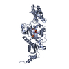
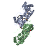
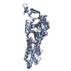
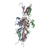
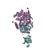
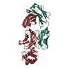
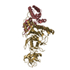
A: DNA polymerase subunit delta-2 B: DNA polymerase subunit delta-3 C: DNA polymerase subunit delta-2 D: DNA polymerase subunit delta-3 E: DNA polymerase subunit delta-2 F: DNA polymerase subunit delta-3 G: DNA polymerase subunit delta-2 H: DNA polymerase subunit delta-3
DNApolymerasesubunitdelta-2 / DNA polymerase subunit delta p50
Mass: 52224.102 Da / Num. of mol.: 4 Source method: isolated from a genetically manipulated source Source: (gene. exp.) Homo sapiens (human) / References: UniProt: P49005, DNA-directed DNA polymerase
#2: Protein
DNApolymerasesubunitdelta-3 / DNA polymerase subunit delta p66
Mass: 16233.576 Da / Num. of mol.: 4 / Fragment: residues 1-144 Source method: isolated from a genetically manipulated source Source: (gene. exp.) Homo sapiens (human) / References: UniProt: Q15054
In the structure databanks used in Yorodumi, some data are registered as the other names, "COVID-19 virus" and "2019-nCoV". Here are the details of the virus and the list of structure data.
Jan 31, 2019. EMDB accession codes are about to change! (news from PDBe EMDB page)
EMDB accession codes are about to change! (news from PDBe EMDB page)
The allocation of 4 digits for EMDB accession codes will soon come to an end. Whilst these codes will remain in use, new EMDB accession codes will include an additional digit and will expand incrementally as the available range of codes is exhausted. The current 4-digit format prefixed with “EMD-” (i.e. EMD-XXXX) will advance to a 5-digit format (i.e. EMD-XXXXX), and so on. It is currently estimated that the 4-digit codes will be depleted around Spring 2019, at which point the 5-digit format will come into force.
The EM Navigator/Yorodumi systems omit the EMD- prefix.
Related info.:Q: What is EMD? / ID/Accession-code notation in Yorodumi/EM Navigator
Yorodumi is a browser for structure data from EMDB, PDB, SASBDB, etc.
This page is also the successor to EM Navigator detail page, and also detail information page/front-end page for Omokage search.
The word "yorodu" (or yorozu) is an old Japanese word meaning "ten thousand". "mi" (miru) is to see.
Related info.:EMDB / PDB / SASBDB / Comparison of 3 databanks / Yorodumi Search / Aug 31, 2016. New EM Navigator & Yorodumi / Yorodumi Papers / Jmol/JSmol / Function and homology information / Changes in new EM Navigator and Yorodumi
 Movie
Movie Controller

 Yorodumi
 Open data
 Basic information
 Components
 Keywords
 Function and homology information
 Homo sapiens (human)
 X-RAY DIFFRACTION /
  Authors
 Citation
 Structure visualization
 Downloads & links
 Other downloads


 PDBj
PDBj


 Assembly
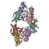

 Mass: 18.015 Da / Num. of mol.: 61 / Source method: isolated from a natural source / Formula: H2O
 Sample preparation
 Processing