 Movie
Movie Controller

[English] 日本語
 Yorodumi
- PDB-1xdm: Structure of human aldolase B associated with hereditary fructose... -
+ Open data

- Basic information
| Entry | Database: PDB / ID: 1xdm | ||||||
|---|---|---|---|---|---|---|---|
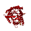
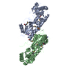
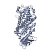
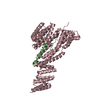
| Title | Structure of human aldolase B associated with hereditary fructose intolerance (A149P), at 291K | ||||||
 Components Components | Fructose-bisphosphate aldolase B | ||||||
 Keywords Keywords | LYASE / ALPHA/BETA BARREL | ||||||
| Function / homology |  Function and homology information Function and homology informationHereditary fructose intolerance / fructose-1-phosphate aldolase activity / : / vacuolar proton-transporting V-type ATPase complex assembly / fructose binding / Fructose catabolism / fructose-bisphosphate aldolase / fructose-bisphosphate aldolase activity / fructose 1,6-bisphosphate metabolic process / fructose metabolic process ...Hereditary fructose intolerance / fructose-1-phosphate aldolase activity / : / vacuolar proton-transporting V-type ATPase complex assembly / fructose binding / Fructose catabolism / fructose-bisphosphate aldolase / fructose-bisphosphate aldolase activity / fructose 1,6-bisphosphate metabolic process / fructose metabolic process / Gluconeogenesis / negative regulation of pentose-phosphate shunt / Glycolysis / microtubule organizing center / cytoskeletal protein binding / centriolar satellite / gluconeogenesis / glycolytic process / ATPase binding / molecular adaptor activity / extracellular exosome / identical protein binding / cytosol Similarity search - Function | ||||||
| Biological species |  Homo sapiens (human) Homo sapiens (human) | ||||||
| Method |  X-RAY DIFFRACTION / MOLECULAR REPLACEMENT / Resolution: 3 Å X-RAY DIFFRACTION / MOLECULAR REPLACEMENT / Resolution: 3 Å | ||||||
 Authors Authors | Malay, A.D. / Allen, K.N. / Tolan, D.R. | ||||||
 Citation Citation | Journal: J.Mol.Biol. / Year: 2005 Title: Structure of the thermolabile mutant aldolase B, A149P: molecular basis of hereditary fructose intolerance. Authors: Malay, A.D. / Allen, K.N. / Tolan, D.R. #1: Journal: Arch.Biochem.Biophys. / Year: 2002Title: The temperature dependence of activity and structure for the most prevalent mutant aldolase B associated with hereditary fructose intolerance Authors: Malay, A.D. / Procious, S.L. / Tolan, D.R. | ||||||
| History |
|
- Structure visualization
| Structure viewer | Molecule: MolmilJmol/JSmol |
|---|
- Downloads & links
-Download
| PDBx/mmCIF format | 1xdm.cif.gz | 423.9 KB | Display | PDBx/mmCIF format |
|---|---|---|---|---|
| PDB format | pdb1xdm.ent.gz | 352.7 KB | Display | PDB format |
| PDBx/mmJSON format | 1xdm.json.gz | Tree view | PDBx/mmJSON format | |
| Others |  Other downloads Other downloads |
-Validation report
| Arichive directory | https://data.pdbj.org/pub/pdb/validation_reports/xd/1xdmftp://data.pdbj.org/pub/pdb/validation_reports/xd/1xdm | HTTPS FTP |
|---|
-Related structure data
| Related structure data | 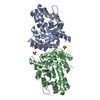 1xdlC 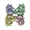 1qo5S C: citing same article ( S: Starting model for refinement |
|---|---|
| Similar structure data |


-Links
 PDBj
PDBj

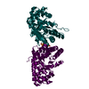
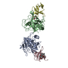
- Assembly
| Deposited unit | 
| ||||||||
|---|---|---|---|---|---|---|---|---|---|
| 1 | 
| ||||||||
| 2 | 
| ||||||||
| 3 | 
| ||||||||
| 4 | 
| ||||||||
| Unit cell |
| ||||||||
| Details | The biological unit of AP-aldolase is a dimer, corresponding to monomer pairs AB, CD, WX, and YZ. The wild-type enzyme is tetrameric. |
-Components
| #1: Protein | Mass: 39558.938 Da / Num. of mol.: 8 / Mutation: A149P Source method: isolated from a genetically manipulated source Source: (gene. exp.) Homo sapiens (human) / Gene: ALDOB, ALDB / Plasmid: pGEX-AP / Production host:  #2: Chemical | ChemComp-SO4 /   Mass: 96.063 Da / Num. of mol.: 8 / Source method: obtained synthetically / Formula: SO4 Mass: 96.063 Da / Num. of mol.: 8 / Source method: obtained synthetically / Formula: SO4#3: Water | ChemComp-HOH / |  Mass: 18.015 Da / Num. of mol.: 35 / Source method: isolated from a natural source / Formula: H2O Mass: 18.015 Da / Num. of mol.: 35 / Source method: isolated from a natural source / Formula: H2O |
|---|
-Experimental details
-Experiment
| Experiment | Method: X-RAY DIFFRACTION / Number of used crystals: 1 |
|---|
- Sample preparation
| Crystal | Density Matthews: 3.4 Å3/Da / Density % sol: 63.7 % |
|---|---|
| Crystal grow | Temperature: 291 K / Method: vapor diffusion, hanging drop / pH: 7.4 Details: ammonium sulfate, 1,8-diaminooctane, pH 7.4, VAPOR DIFFUSION, HANGING DROP, temperature 291K |
-Data collection
| Diffraction | Mean temperature: 93 K |
|---|---|
| Diffraction source | Source: ROTATING ANODE / Type: RIGAKU RU300 / Wavelength: 1.5418 Å |
| Detector | Type: RIGAKU RAXIS IV / Detector: IMAGE PLATE / Date: Jan 13, 2003 |
| Radiation | Monochromator: GRAPHITE / Protocol: SINGLE WAVELENGTH / Monochromatic (M) / Laue (L): M / Scattering type: x-ray |
| Radiation wavelength | Wavelength: 1.5418 Å / Relative weight: 1 |
| Reflection | Resolution: 3→100 Å / Num. all: 87953 / Num. obs: 83595 / % possible obs: 95.1 % / Observed criterion σ(I): 0 / Redundancy: 4.4 % / Rsym value: 0.067 / Net I/σ(I): 12.1 |
| Reflection shell | Resolution: 3→3.11 Å / Redundancy: 2.5 % / Mean I/σ(I) obs: 2.1 / Num. unique all: 7680 / Rsym value: 0.364 / % possible all: 88.8 |
- Processing
| Software |
| ||||||||||||||||||||||||||||||||||||||||||||||||||||||||||||
|---|---|---|---|---|---|---|---|---|---|---|---|---|---|---|---|---|---|---|---|---|---|---|---|---|---|---|---|---|---|---|---|---|---|---|---|---|---|---|---|---|---|---|---|---|---|---|---|---|---|---|---|---|---|---|---|---|---|---|---|---|---|
| Refinement | Method to determine structure: MOLECULAR REPLACEMENT Starting model: PDB entry 1QO5 Resolution: 3→79.4 Å / Rfactor Rfree error: 0.004 / Data cutoff high absF: 295432.84 / Data cutoff low absF: 0 / Isotropic thermal model: GROUP / Cross valid method: THROUGHOUT / σ(F): 2 / Stereochemistry target values: Engh & Huber / Details: used ncs restraints between monomers
| ||||||||||||||||||||||||||||||||||||||||||||||||||||||||||||
| Solvent computation | Solvent model: FLAT MODEL / Bsol: 13.4177 Å2 / ksol: 0.294477 e/Å3 | ||||||||||||||||||||||||||||||||||||||||||||||||||||||||||||
| Displacement parameters | Biso mean: 60.3 Å2
| ||||||||||||||||||||||||||||||||||||||||||||||||||||||||||||
| Refine analyze |
| ||||||||||||||||||||||||||||||||||||||||||||||||||||||||||||
| Refinement step | Cycle: LAST / Resolution: 3→79.4 Å
| ||||||||||||||||||||||||||||||||||||||||||||||||||||||||||||
| Refine LS restraints |
| ||||||||||||||||||||||||||||||||||||||||||||||||||||||||||||
| LS refinement shell | Resolution: 3→3.11 Å / Rfactor Rfree error: 0.018 / Total num. of bins used: 10
| ||||||||||||||||||||||||||||||||||||||||||||||||||||||||||||
| Xplor file |
|
