 Movie
Movie Controller

[English] 日本語
 Yorodumi
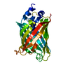
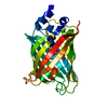
- PDB-3dpz: Structure of the Yellow Fluorescent Protein Citrine Frozen at 400... -
+ Open data

- Basic information
| Entry | Database: PDB / ID: 3dpz | ||||||
|---|---|---|---|---|---|---|---|
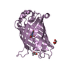
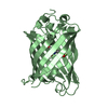
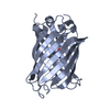
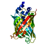
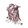
| Title | Structure of the Yellow Fluorescent Protein Citrine Frozen at 4000 Atmospheres Number 3: Structure 25 in a Series of 26 High Pressure Structures | ||||||
 Components Components | Green fluorescent protein | ||||||
 Keywords Keywords | LUMINESCENT PROTEIN / Yellow Fluorescent Protein / beta barrel / chromophore / fluorescent protein / high pressure / Luminescence / Photoprotein | ||||||
| Function / homology |  Function and homology information Function and homology information | ||||||
| Biological species |   Aequorea victoria (jellyfish) Aequorea victoria (jellyfish) | ||||||
| Method | X-RAY DIFFRACTION / SYNCHROTRON / MOLECULAR REPLACEMENT / molecular replacement / Resolution: 1.7 Å | ||||||
 Authors Authors | Barstow, B. / Kim, C.U. | ||||||
 Citation Citation | Journal: Proc.Natl.Acad.Sci.Usa / Year: 2008 Title: Alteration of citrine structure by hydrostatic pressure explains the accompanying spectral shift. Authors: Barstow, B. / Ando, N. / Kim, C.U. / Gruner, S.M. #1: Journal: Acta Crystallogr.,Sect.D / Year: 2005 Title: High-pressure cooling of protein crystals without cryoprotectants. Authors: Kim, C.U. / Kapfer, R. / Gruner, S.M. | ||||||
| History |
|
- Structure visualization
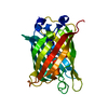
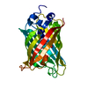
| Structure viewer | Molecule: MolmilJmol/JSmol |
|---|
- Downloads & links
-Download
| PDBx/mmCIF format | 3dpz.cif.gz | 67 KB | Display | PDBx/mmCIF format |
|---|---|---|---|---|
| PDB format | pdb3dpz.ent.gz | 47.4 KB | Display | PDB format |
| PDBx/mmJSON format | 3dpz.json.gz | Tree view | PDBx/mmJSON format | |
| Others |  Other downloads Other downloads |
-Validation report
| Arichive directory | https://data.pdbj.org/pub/pdb/validation_reports/dp/3dpzftp://data.pdbj.org/pub/pdb/validation_reports/dp/3dpz | HTTPS FTP |
|---|
-Related structure data
| Related structure data |  3dpwC  3dpxC  3dq1C  3dq2C  3dq3C  3dq4C  3dq5C  3dq6C 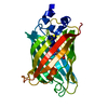 3dq7C  3dq8C  3dq9C  3dqaC  3dqcC  3dqdC  3dqeC  3dqfC  3dqhC  3dqiC  3dqjC  3dqkC  3dqlC  3dqmC  3dqnC  3dqoC 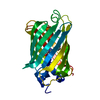 3dquC  1huyS S: Starting model for refinement C: citing same article ( |
|---|---|
| Similar structure data |


-Links
 PDBj
PDBj

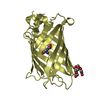
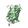
- Assembly
| Deposited unit | 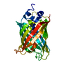
| ||||||||
|---|---|---|---|---|---|---|---|---|---|
| 1 |
| ||||||||
| Unit cell |
|
-Components
| #1: Protein | Mass: 27393.916 Da / Num. of mol.: 1 / Mutation: S65G, V68L, Q69M, S72A, T203Y Source method: isolated from a genetically manipulated source Source: (gene. exp.) Aequorea victoria (jellyfish) / Gene: GFP / Plasmid: pET28 / Production host:  Escherichia coli (E. coli) / Strain (production host): BL21(DE3) / References: UniProt: P42212 Escherichia coli (E. coli) / Strain (production host): BL21(DE3) / References: UniProt: P42212 |
|---|---|
| #2: Water | ChemComp-HOH / Water Mass: 18.015 Da / Num. of mol.: 205 / Source method: isolated from a natural source / Formula: H2O Mass: 18.015 Da / Num. of mol.: 205 / Source method: isolated from a natural source / Formula: H2O |
| Sequence details | RESIDUE SER 65 HAS BEEN MUTATED TO GLY 65. RESIDUES GLY 65, TYR 66 AND GLY 67 CONSTITUTE THE ...RESIDUE SER 65 HAS BEEN MUTATED TO GLY 65. RESIDUES GLY 65, TYR 66 AND GLY 67 CONSTITUTE |
-Experimental details
-Experiment
| Experiment | Method: X-RAY DIFFRACTION / Number of used crystals: 1 |
|---|
- Sample preparation
| Crystal | Density Matthews: 2.11 Å3/Da / Density % sol: 41.59 % Description: Crystal structure of the Yellow Fluorescent Protein Citrine frozen at 4000 atmospheres. Structure 25 of 26 in a series of high pressure structures. Crystal was high pressure cryo-cooled ...Description: Crystal structure of the Yellow Fluorescent Protein Citrine frozen at 4000 atmospheres. Structure 25 of 26 in a series of high pressure structures. Crystal was high pressure cryo-cooled at 4000 atmospheres in helium gas. Crystal temperature was maintained below 100 K prior to data collection at ambient pressure and 100 K. High pressure cryo-cooling procedure is described in secondary citation 1 (Kim et al., Acta Cryst. D61:881-890). Structure referred to as citrine4000_3 in primary citation (Barstow et al.). |
|---|---|
| Crystal grow | Temperature: 277 K / Method: vapor diffusion, hanging drop / pH: 5 Details: Crystals grown by seeding using a Seed Bead (HR2-320, Hampton Research) in 5% PEG 3350, 50 mM Na Acetate, 50 mM NH4 Acetate, pH 5.0. Crystals were grown at 4 deg C and at ambient pressure, ...Details: Crystals grown by seeding using a Seed Bead (HR2-320, Hampton Research) in 5% PEG 3350, 50 mM Na Acetate, 50 mM NH4 Acetate, pH 5.0. Crystals were grown at 4 deg C and at ambient pressure, VAPOR DIFFUSION, HANGING DROP, temperature 277K |
-Data collection
| Diffraction | Mean temperature: 100 K |
|---|---|
| Diffraction source | Source: SYNCHROTRON / Site: CHESS  / Beamline: F2 / Wavelength: 0.9795 Å / Beamline: F2 / Wavelength: 0.9795 Å |
| Detector | Type: ADSC QUANTUM 210 / Detector: CCD / Date: Apr 5, 2007 |
| Radiation | Monochromator: Si(111) DOUBLE CRYSTAL / Protocol: SINGLE WAVELENGTH / Monochromatic (M) / Laue (L): M / Scattering type: x-ray |
| Radiation wavelength | Wavelength: 0.9795 Å / Relative weight: 1 |
| Reflection | Resolution: 1.7→41.81 Å / Num. obs: 25414 / % possible obs: 97.4 % / Redundancy: 6.6 % / Rmerge(I) obs: 0.069 / Rsym value: 0.069 / Net I/σ(I): 8.6 |
| Reflection shell | Highest resolution: 1.7 Å / Redundancy: 6.8 % / Rmerge(I) obs: 0.259 / Mean I/σ(I) obs: 2.9 / Rsym value: 0.259 / % possible all: 99.9 |
-Phasing
| Phasing | Method: molecular replacement | |||||||||
|---|---|---|---|---|---|---|---|---|---|---|
| Phasing MR |
|
- Processing
| Software |
| ||||||||||||||||||||||||||||||||||||||||||||||||||||||||||||||||||||||||||||||||||||||||||||||||||||||||||||||||||||||||||||||||||||||||||||||||||||||||||||||||||||||||||
|---|---|---|---|---|---|---|---|---|---|---|---|---|---|---|---|---|---|---|---|---|---|---|---|---|---|---|---|---|---|---|---|---|---|---|---|---|---|---|---|---|---|---|---|---|---|---|---|---|---|---|---|---|---|---|---|---|---|---|---|---|---|---|---|---|---|---|---|---|---|---|---|---|---|---|---|---|---|---|---|---|---|---|---|---|---|---|---|---|---|---|---|---|---|---|---|---|---|---|---|---|---|---|---|---|---|---|---|---|---|---|---|---|---|---|---|---|---|---|---|---|---|---|---|---|---|---|---|---|---|---|---|---|---|---|---|---|---|---|---|---|---|---|---|---|---|---|---|---|---|---|---|---|---|---|---|---|---|---|---|---|---|---|---|---|---|---|---|---|---|---|---|
| Refinement | Method to determine structure: MOLECULAR REPLACEMENT Starting model: PDB entry 1HUY Resolution: 1.7→20 Å / Cor.coef. Fo:Fc: 0.94 / Cor.coef. Fo:Fc free: 0.877 / Occupancy max: 1 / Occupancy min: 1 / SU B: 3.082 / SU ML: 0.103 / Cross valid method: THROUGHOUT / ESU R: 0.138 / ESU R Free: 0.15 / Stereochemistry target values: MAXIMUM LIKELIHOOD Details: 1. HYDROGENS HAVE BEEN ADDED IN THE RIDING POSITIONS. 2. This structure is refined slightly differently from the corresponding structure used for analysis in the primary citation (Barstow et ...Details: 1. HYDROGENS HAVE BEEN ADDED IN THE RIDING POSITIONS. 2. This structure is refined slightly differently from the corresponding structure used for analysis in the primary citation (Barstow et al.). However, analysis of the deformation motion of the chromophore under pressure in this sequence of deposited structures produces an identical deformation trend. For copies of the structures as used in the analysis in the primary citation please contact the authors.
| ||||||||||||||||||||||||||||||||||||||||||||||||||||||||||||||||||||||||||||||||||||||||||||||||||||||||||||||||||||||||||||||||||||||||||||||||||||||||||||||||||||||||||
| Solvent computation | Ion probe radii: 0.8 Å / Shrinkage radii: 0.8 Å / VDW probe radii: 1.2 Å / Solvent model: MASK | ||||||||||||||||||||||||||||||||||||||||||||||||||||||||||||||||||||||||||||||||||||||||||||||||||||||||||||||||||||||||||||||||||||||||||||||||||||||||||||||||||||||||||
| Displacement parameters | Biso mean: 23.531 Å2
| ||||||||||||||||||||||||||||||||||||||||||||||||||||||||||||||||||||||||||||||||||||||||||||||||||||||||||||||||||||||||||||||||||||||||||||||||||||||||||||||||||||||||||
| Refinement step | Cycle: LAST / Resolution: 1.7→20 Å
| ||||||||||||||||||||||||||||||||||||||||||||||||||||||||||||||||||||||||||||||||||||||||||||||||||||||||||||||||||||||||||||||||||||||||||||||||||||||||||||||||||||||||||
| Refine LS restraints |
| ||||||||||||||||||||||||||||||||||||||||||||||||||||||||||||||||||||||||||||||||||||||||||||||||||||||||||||||||||||||||||||||||||||||||||||||||||||||||||||||||||||||||||
| LS refinement shell | Resolution: 1.7→1.744 Å / Total num. of bins used: 20
|
