 Movie
Movie Controller

[English] 日本語
 Yorodumi
- PDB-4ryw: Crystal structure of the photoconverted green fluorescent protein... -
+ Open data

- Basic information
| Entry | Database: PDB / ID: 4ryw | ||||||
|---|---|---|---|---|---|---|---|
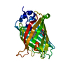
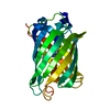
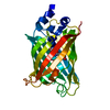
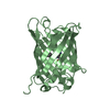
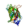
| Title | Crystal structure of the photoconverted green fluorescent protein NowGFP_conv (the variant of cyan Cerulean) at pH 7.0 | ||||||
 Components Components | NowGFP_conv | ||||||
 Keywords Keywords | FLUORESCENT PROTEIN / photoconverted NowGFP / TWG chromophore / beta-barrel / variant of cyan Cerulean | ||||||
| Function / homology | Green Fluorescent Protein / Green fluorescent protein / Beta Barrel / Mainly Beta Function and homology information Function and homology information | ||||||
| Method |  X-RAY DIFFRACTION / SYNCHROTRON / MOLECULAR REPLACEMENT / Resolution: 2.5 Å X-RAY DIFFRACTION / SYNCHROTRON / MOLECULAR REPLACEMENT / Resolution: 2.5 Å | ||||||
 Authors Authors | Pletnev, V.Z. / Pletneva, N.V. / Pletnev, S.V. | ||||||
 Citation Citation | Journal: Acta Crystallogr.,Sect.D / Year: 2015 Title: Structure of the green fluorescent protein NowGFP with an anionic tryptophan-based chromophore. Authors: Pletnev, V.Z. / Pletneva, N.V. / Sarkisyan, K.S. / Mishin, A.S. / Lukyanov, K.A. / Goryacheva, E.A. / Ziganshin, R.H. / Dauter, Z. / Pletnev, S. | ||||||
| History |
|
- Structure visualization
| Structure viewer | Molecule: MolmilJmol/JSmol |
|---|
- Downloads & links
-Download
| PDBx/mmCIF format | 4ryw.cif.gz | 150.1 KB | Display | PDBx/mmCIF format |
|---|---|---|---|---|
| PDB format | pdb4ryw.ent.gz | 118.5 KB | Display | PDB format |
| PDBx/mmJSON format | 4ryw.json.gz | Tree view | PDBx/mmJSON format | |
| Others |  Other downloads Other downloads |
-Validation report
| Arichive directory | https://data.pdbj.org/pub/pdb/validation_reports/ry/4rywftp://data.pdbj.org/pub/pdb/validation_reports/ry/4ryw | HTTPS FTP |
|---|
-Related structure data
| Related structure data |  4rtcC  4rysC 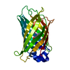 2wsoS S: Starting model for refinement C: citing same article ( |
|---|---|
| Similar structure data |


-Links
 PDBj
PDBj- Assembly
| Deposited unit | 
| ||||||||
|---|---|---|---|---|---|---|---|---|---|
| 1 | 
| ||||||||
| 2 | 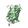
| ||||||||
| 3 | 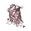
| ||||||||
| Unit cell |
| ||||||||
| Details | Three molecules in the assymetric unit |
-Components
| #1: Protein | Mass: 27969.389 Da / Num. of mol.: 3 Source method: isolated from a genetically manipulated source Production host:  #2: Chemical | ChemComp-GOL /   Mass: 92.094 Da / Num. of mol.: 9 / Source method: obtained synthetically / Formula: C3H8O3 Mass: 92.094 Da / Num. of mol.: 9 / Source method: obtained synthetically / Formula: C3H8O3#3: Water | ChemComp-HOH / |  Mass: 18.015 Da / Num. of mol.: 164 / Source method: isolated from a natural source / Formula: H2O Mass: 18.015 Da / Num. of mol.: 164 / Source method: isolated from a natural source / Formula: H2OHas protein modification | Y | Sequence details | This construct is a gene engineered variant of the cyan fluorescent protein Cerulean | |
|---|
-Experimental details
-Experiment
| Experiment | Method: X-RAY DIFFRACTION / Number of used crystals: 1 |
|---|
- Sample preparation
| Crystal | Density Matthews: 2.03 Å3/Da / Density % sol: 39.37 % |
|---|---|
| Crystal grow | Temperature: 293 K / Method: vapor diffusion, hanging drop / pH: 7 Details: 0.1M Na Acetate pH=4.5, 30% (v/v) PEG 5000 mono methyl ether, pH 7.0, VAPOR DIFFUSION, HANGING DROP, temperature 293.0K |
-Data collection
| Diffraction | Mean temperature: 100 K | |||||||||||||||||||||||||||||||||||||||||||||||||||||||||||||||||||||||||||||
|---|---|---|---|---|---|---|---|---|---|---|---|---|---|---|---|---|---|---|---|---|---|---|---|---|---|---|---|---|---|---|---|---|---|---|---|---|---|---|---|---|---|---|---|---|---|---|---|---|---|---|---|---|---|---|---|---|---|---|---|---|---|---|---|---|---|---|---|---|---|---|---|---|---|---|---|---|---|---|
| Diffraction source | Source: SYNCHROTRON / Site: APS  / Beamline: 22-ID / Wavelength: 1 Å / Beamline: 22-ID / Wavelength: 1 Å | |||||||||||||||||||||||||||||||||||||||||||||||||||||||||||||||||||||||||||||
| Detector | Type: MARMOSAIC 300 mm CCD / Detector: CCD / Date: 2014 / Details: mirrors | |||||||||||||||||||||||||||||||||||||||||||||||||||||||||||||||||||||||||||||
| Radiation | Monochromator: mirror / Protocol: SINGLE WAVELENGTH / Monochromatic (M) / Laue (L): M / Scattering type: x-ray | |||||||||||||||||||||||||||||||||||||||||||||||||||||||||||||||||||||||||||||
| Radiation wavelength | Wavelength: 1 Å / Relative weight: 1 | |||||||||||||||||||||||||||||||||||||||||||||||||||||||||||||||||||||||||||||
| Reflection | Resolution: 2.5→30 Å / Num. obs: 23175 / % possible obs: 100 % / Redundancy: 3.7 % / Rmerge(I) obs: 0.117 / Χ2: 0.856 / Net I/σ(I): 6.6 | |||||||||||||||||||||||||||||||||||||||||||||||||||||||||||||||||||||||||||||
| Reflection shell |
|
- Processing
| Software |
| |||||||||||||||||||||||||||||||||||||||||||||||||||||||||||||||||||||||||||
|---|---|---|---|---|---|---|---|---|---|---|---|---|---|---|---|---|---|---|---|---|---|---|---|---|---|---|---|---|---|---|---|---|---|---|---|---|---|---|---|---|---|---|---|---|---|---|---|---|---|---|---|---|---|---|---|---|---|---|---|---|---|---|---|---|---|---|---|---|---|---|---|---|---|---|---|---|
| Refinement | Method to determine structure: MOLECULAR REPLACEMENT Starting model: PDB ENTRY 2WSO Resolution: 2.5→30 Å / Cor.coef. Fo:Fc: 0.922 / Cor.coef. Fo:Fc free: 0.841 / WRfactor Rfree: 0.2862 / WRfactor Rwork: 0.2033 / FOM work R set: 0.769 / SU B: 13.06 / SU ML: 0.297 / SU Rfree: 0.3815 / Cross valid method: THROUGHOUT / σ(F): 0 / ESU R Free: 0.384 / Stereochemistry target values: MAXIMUM LIKELIHOOD Details: HYDROGENS HAVE BEEN ADDED IN THE RIDING POSITIONS U VALUES : REFINED INDIVIDUALLY
| |||||||||||||||||||||||||||||||||||||||||||||||||||||||||||||||||||||||||||
| Solvent computation | Ion probe radii: 0.8 Å / Shrinkage radii: 0.8 Å / VDW probe radii: 1.2 Å / Solvent model: MASK | |||||||||||||||||||||||||||||||||||||||||||||||||||||||||||||||||||||||||||
| Displacement parameters | Biso max: 92.95 Å2 / Biso mean: 37.682 Å2 / Biso min: 14.35 Å2
| |||||||||||||||||||||||||||||||||||||||||||||||||||||||||||||||||||||||||||
| Refinement step | Cycle: LAST / Resolution: 2.5→30 Å
| |||||||||||||||||||||||||||||||||||||||||||||||||||||||||||||||||||||||||||
| Refine LS restraints |
| |||||||||||||||||||||||||||||||||||||||||||||||||||||||||||||||||||||||||||
| LS refinement shell | Resolution: 2.499→2.564 Å / Total num. of bins used: 20
|
