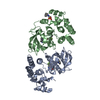
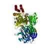
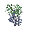
ERBB2 signaling pathway / negative regulation of beige fat cell differentiation / reelin-mediated signaling pathway / cullin-RING-type E3 NEDD8 transferase / NEDD8 transferase activity / regulation of neuron migration / cullin-RING ubiquitin ligase complex / Cul7-RING ubiquitin ligase complex / cellular response to chemical stress / Loss of Function of FBXW7 in Cancer and NOTCH1 Signaling ...ERBB2 signaling pathway / negative regulation of beige fat cell differentiation / reelin-mediated signaling pathway / cullin-RING-type E3 NEDD8 transferase / NEDD8 transferase activity / regulation of neuron migration / cullin-RING ubiquitin ligase complex / Cul7-RING ubiquitin ligase complex / cellular response to chemical stress / Loss of Function of FBXW7 in Cancer and NOTCH1 Signaling / protein K27-linked ubiquitination / positive regulation of protein autoubiquitination / RNA polymerase II transcription initiation surveillance / protein K11-linked ubiquitination / protein neddylation / NEDD8 ligase activity / VCB complex / negative regulation of response to oxidative stress / Cul5-RING ubiquitin ligase complex / SCF ubiquitin ligase complex / ubiquitin-ubiquitin ligase activity / ubiquitin-dependent protein catabolic process via the C-end degron rule pathway / Cul2-RING ubiquitin ligase complex / SCF-dependent proteasomal ubiquitin-dependent protein catabolic process / negative regulation of type I interferon production / Cul3-RING ubiquitin ligase complex / negative regulation of mitophagy / Cul4A-RING E3 ubiquitin ligase complex / Cul4-RING E3 ubiquitin ligase complex / Prolactin receptor signaling / Cul4B-RING E3 ubiquitin ligase complex / ubiquitin ligase complex scaffold activity / cullin family protein binding / protein monoubiquitination / site of DNA damage / protein K48-linked ubiquitination / signal transduction in response to DNA damage / Nuclear events stimulated by ALK signaling in cancer / transcription-coupled nucleotide-excision repair / positive regulation of TORC1 signaling / regulation of cellular response to insulin stimulus / negative regulation of insulin receptor signaling pathway / intrinsic apoptotic signaling pathway / post-translational protein modification / negative regulation of canonical NF-kappaB signal transduction / T cell activation / Regulation of BACH1 activity / Degradation of CRY and PER proteins / cellular response to amino acid stimulus / Degradation of DVL / G1/S transition of mitotic cell cycle / Degradation of GLI1 by the proteasome / negative regulation of canonical Wnt signaling pathway / GSK3B and BTRC:CUL1-mediated-degradation of NFE2L2 / Negative regulation of NOTCH4 signaling / Ubiquitin-Mediated Degradation of Phosphorylated Cdc25A / Recognition of DNA damage by PCNA-containing replication complex / Hedgehog 'on' state / Vif-mediated degradation of APOBEC3G / FBXL7 down-regulates AURKA during mitotic entry and in early mitosis / Degradation of GLI2 by the proteasome / GLI3 is processed to GLI3R by the proteasome / Inactivation of CSF3 (G-CSF) signaling / RING-type E3 ubiquitin transferase / Degradation of beta-catenin by the destruction complex / Oxygen-dependent proline hydroxylation of Hypoxia-inducible Factor Alpha / DNA Damage Recognition in GG-NER / Evasion by RSV of host interferon responses / NOTCH1 Intracellular Domain Regulates Transcription / Constitutive Signaling by NOTCH1 PEST Domain Mutants / Constitutive Signaling by NOTCH1 HD+PEST Domain Mutants / Dual Incision in GG-NER / Transcription-Coupled Nucleotide Excision Repair (TC-NER) / calcium channel activity / Formation of TC-NER Pre-Incision Complex / Downregulation of ERBB2 signaling / Regulation of expression of SLITs and ROBOs / Formation of Incision Complex in GG-NER / Interleukin-1 signaling / protein polyubiquitination / Orc1 removal from chromatin / Regulation of RAS by GAPs / Dual incision in TC-NER / Gap-filling DNA repair synthesis and ligation in TC-NER / Regulation of RUNX2 expression and activity / positive regulation of protein catabolic process / ubiquitin-protein transferase activity / cellular response to UV / ubiquitin protein ligase activity / KEAP1-NFE2L2 pathway / Antigen processing: Ubiquitination & Proteasome degradation / MAPK cascade / positive regulation of proteasomal ubiquitin-dependent protein catabolic process / signaling receptor activity / Neddylation / cellular response to oxidative stress / spermatogenesis / protein-macromolecule adaptor activity / ubiquitin-dependent protein catabolic process / Potential therapeutics for SARS Similarity search - Function
Type: ADSC QUANTUM 315 / Detector: CCD / Date: Nov 8, 2006 Details: CRYOGENICALLY COOLED FIRST CRYSTAL AND SAGITALLY BENT SECOND CRYSTAL HORIZONTALLY FOCUSING
Radiation
Monochromator: SI(111) DOUBLE CRYSTAL / Protocol: SINGLE WAVELENGTH / Monochromatic (M) / Laue (L): M / Scattering type: x-ray
Radiation wavelength
Wavelength: 1 Å / Relative weight: 1
Reflection
Resolution: 2.55→50 Å / Num. obs: 18719 / % possible obs: 95.5 % / Redundancy: 5.2 % / Biso Wilson estimate: 37.8 Å2 / Rmerge(I) obs: 0.156 / Net I/σ(I): 16.1
Reflection shell
Resolution: 2.55→2.64 Å / Redundancy: 2.9 % / Rmerge(I) obs: 0.407 / Mean I/σ(I) obs: 2
-
Processing
Software
Name
Version
Classification
ADSC
Quantum
datacollection
CNS
refinement
HKL-2000
datareduction
HKL-2000
datascaling
CNS
phasing
Refinement
Method to determine structure: MOLECULAR REPLACEMENT / Resolution: 2.6→50 Å / Rfactor Rfree error: 0.009 / Data cutoff high absF: 1323701.74 / Data cutoff low absF: 0 / Isotropic thermal model: RESTRAINED / Cross valid method: THROUGHOUT / σ(F): 0
In the structure databanks used in Yorodumi, some data are registered as the other names, "COVID-19 virus" and "2019-nCoV". Here are the details of the virus and the list of structure data.
Jan 31, 2019. EMDB accession codes are about to change! (news from PDBe EMDB page)
EMDB accession codes are about to change! (news from PDBe EMDB page)
The allocation of 4 digits for EMDB accession codes will soon come to an end. Whilst these codes will remain in use, new EMDB accession codes will include an additional digit and will expand incrementally as the available range of codes is exhausted. The current 4-digit format prefixed with “EMD-” (i.e. EMD-XXXX) will advance to a 5-digit format (i.e. EMD-XXXXX), and so on. It is currently estimated that the 4-digit codes will be depleted around Spring 2019, at which point the 5-digit format will come into force.
The EM Navigator/Yorodumi systems omit the EMD- prefix.
Related info.:Q: What is EMD? / ID/Accession-code notation in Yorodumi/EM Navigator
Yorodumi is a browser for structure data from EMDB, PDB, SASBDB, etc.
This page is also the successor to EM Navigator detail page, and also detail information page/front-end page for Omokage search.
The word "yorodu" (or yorozu) is an old Japanese word meaning "ten thousand". "mi" (miru) is to see.
Related info.:EMDB / PDB / SASBDB / Comparison of 3 databanks / Yorodumi Search / Aug 31, 2016. New EM Navigator & Yorodumi / Yorodumi Papers / Jmol/JSmol / Function and homology information / Changes in new EM Navigator and Yorodumi
 Movie
Movie Controller

 Yorodumi
 Open data
 Basic information
 Components
 Keywords
 Function and homology information
 Homo sapiens (human)
 X-RAY DIFFRACTION /
  Authors
 Citation
 Structure visualization
 Downloads & links
 Other downloads
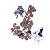


 PDBj
PDBj


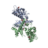
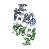
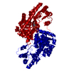
 Assembly


 Mass: 65.409 Da / Num. of mol.: 3 / Source method: obtained synthetically / Formula: Zn
 Mass: 18.015 Da / Num. of mol.: 73 / Source method: isolated from a natural source / Formula: H2O
 Sample preparation
 / Beamline: X25 / Wavelength: 1
  Processing