 Movie
Movie Controller

[English] 日本語
 Yorodumi
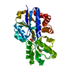
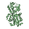
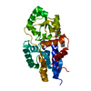
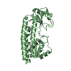
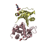
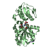
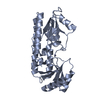
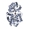
- PDB-3chg: The compatible solute-binding protein OpuAC from Bacillus subtili... -
+ Open data

- Basic information
| Entry | Database: PDB / ID: 3chg | ||||||
|---|---|---|---|---|---|---|---|
| Title | The compatible solute-binding protein OpuAC from Bacillus subtilis in complex with DMSA | ||||||
 Components Components | Glycine betaine-binding protein | ||||||
 Keywords Keywords |  LIGAND BINDING PROTEIN / Transport protein / substrate binding protein LIGAND BINDING PROTEIN / Transport protein / substrate binding protein | ||||||
| Function / homology |  Function and homology information Function and homology informationamino acid transport / response to stimulus / transmembrane transporter activity / ATP-binding cassette (ABC) transporter complex / membrane raftSimilarity search - Function | ||||||
| Biological species |  Bacillus subtilis (bacteria) Bacillus subtilis (bacteria) | ||||||
| Method | X-RAY DIFFRACTION / SYNCHROTRON / MOLECULAR REPLACEMENT / Resolution: 2.8 Å | ||||||
 Authors Authors | Smits, S.H.J. / Hoing, M. / Lecher, J. / Jebbar, M. / Schmitt, L. / Bremer, E. | ||||||
 Citation Citation | Journal: J.Bacteriol. / Year: 2008 Title: The Compatible-Solute-Binding Protein OpuAC from Bacillus subtilis: Ligand Binding, Site-Directed Mutagenesis, and Crystallographic Studies Authors: Smits, S.H.J. / Hoing, M. / Lecher, J. / Jebbar, M. / Schmitt, L. / Bremer, E. | ||||||
| History |
|
- Structure visualization
| Structure viewer | Molecule: MolmilJmol/JSmol |
|---|
- Downloads & links
-Download
| PDBx/mmCIF format | 3chg.cif.gz | 201.4 KB | Display | PDBx/mmCIF format |
|---|---|---|---|---|
| PDB format | pdb3chg.ent.gz | 163.3 KB | Display | PDB format |
| PDBx/mmJSON format | 3chg.json.gz | Tree view | PDBx/mmJSON format | |
| Others |  Other downloads Other downloads |
-Validation report
| Arichive directory | https://data.pdbj.org/pub/pdb/validation_reports/ch/3chgftp://data.pdbj.org/pub/pdb/validation_reports/ch/3chg | HTTPS FTP |
|---|
-Related structure data
| Related structure data | |
|---|---|
| Similar structure data |


-Links
 PDBj
PDBj


- Assembly
| Deposited unit | 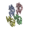
| ||||||||
|---|---|---|---|---|---|---|---|---|---|
| 1 | 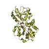
| ||||||||
| 2 | 
| ||||||||
| 3 | 
| ||||||||
| 4 | 
| ||||||||
| Unit cell |
|
-Components
| #1: Protein | Mass: 29780.914 Da / Num. of mol.: 4 Source method: isolated from a genetically manipulated source Source: (gene. exp.) Bacillus subtilis (bacteria) / Gene: opuAC / Plasmid: Pbkb76 OpuAC_wt / Production host: Escherichia coli (E. coli) / Strain (production host): Bl21(DE3) / References: UniProt: P46922#2: Chemical | ChemComp-313 / (   Mass: 122.186 Da / Num. of mol.: 4 / Source method: obtained synthetically / Formula: C4H10O2S Mass: 122.186 Da / Num. of mol.: 4 / Source method: obtained synthetically / Formula: C4H10O2S |
|---|
-Experimental details
-Experiment
| Experiment | Method: X-RAY DIFFRACTION / Number of used crystals: 1 |
|---|
- Sample preparation
| Crystal | Density Matthews: 2.04 Å3/Da / Density % sol: 39.66 % |
|---|---|
| Crystal grow | Temperature: 274 K / Method: vapor diffusion, hanging drop / pH: 8.3 Details: Tris, ammonium acetate, PEG 4000, pH 8.3, VAPOR DIFFUSION, HANGING DROP, temperature 274K |
-Data collection
| Diffraction | Mean temperature: 100 K |
|---|---|
| Diffraction source | Source: SYNCHROTRON / Site: EMBL/DESY, HAMBURG  / Beamline: BW7A / Wavelength: 0.97854 Å / Beamline: BW7A / Wavelength: 0.97854 Å |
| Detector | Type: MAR CCD 165 mm / Detector: CCD / Date: Sep 15, 2007 |
| Radiation | Protocol: SINGLE WAVELENGTH / Monochromatic (M) / Laue (L): M / Scattering type: x-ray |
| Radiation wavelength | Wavelength: 0.97854 Å / Relative weight: 1 |
| Reflection | Resolution: 2.8→20 Å / Num. all: 24818 / Num. obs: 20667 / % possible obs: 93 % / Observed criterion σ(I): 2 / Redundancy: 2.4 % / Rmerge(I) obs: 0.164 |
| Reflection shell | Resolution: 2.8→2.85 Å / Redundancy: 2.2 % / Rmerge(I) obs: 0.29 / Mean I/σ(I) obs: 3 / % possible all: 96.5 |
- Processing
| Software |
| ||||||||||||||||||||||||||||||||||||||||||||||||||||||||||||||||||||||||||||||||||||||||||||||||||||||||||||||||||||||||||||||||||||||||||||||||||||||||||||||||||||||||||
|---|---|---|---|---|---|---|---|---|---|---|---|---|---|---|---|---|---|---|---|---|---|---|---|---|---|---|---|---|---|---|---|---|---|---|---|---|---|---|---|---|---|---|---|---|---|---|---|---|---|---|---|---|---|---|---|---|---|---|---|---|---|---|---|---|---|---|---|---|---|---|---|---|---|---|---|---|---|---|---|---|---|---|---|---|---|---|---|---|---|---|---|---|---|---|---|---|---|---|---|---|---|---|---|---|---|---|---|---|---|---|---|---|---|---|---|---|---|---|---|---|---|---|---|---|---|---|---|---|---|---|---|---|---|---|---|---|---|---|---|---|---|---|---|---|---|---|---|---|---|---|---|---|---|---|---|---|---|---|---|---|---|---|---|---|---|---|---|---|---|---|---|
| Refinement | Method to determine structure: MOLECULAR REPLACEMENT / Resolution: 2.8→19.56 Å / Cor.coef. Fo:Fc: 0.859 / Cor.coef. Fo:Fc free: 0.766 / SU B: 28.413 / SU ML: 0.55 / Cross valid method: THROUGHOUT / ESU R Free: 0.659 / Stereochemistry target values: MAXIMUM LIKELIHOOD / Details: HYDROGENS HAVE BEEN ADDED IN THE RIDING POSITIONS
| ||||||||||||||||||||||||||||||||||||||||||||||||||||||||||||||||||||||||||||||||||||||||||||||||||||||||||||||||||||||||||||||||||||||||||||||||||||||||||||||||||||||||||
| Solvent computation | Ion probe radii: 0.8 Å / Shrinkage radii: 0.8 Å / VDW probe radii: 1.2 Å / Solvent model: MASK | ||||||||||||||||||||||||||||||||||||||||||||||||||||||||||||||||||||||||||||||||||||||||||||||||||||||||||||||||||||||||||||||||||||||||||||||||||||||||||||||||||||||||||
| Displacement parameters | Biso mean: 15.676 Å2
| ||||||||||||||||||||||||||||||||||||||||||||||||||||||||||||||||||||||||||||||||||||||||||||||||||||||||||||||||||||||||||||||||||||||||||||||||||||||||||||||||||||||||||
| Refinement step | Cycle: LAST / Resolution: 2.8→19.56 Å
| ||||||||||||||||||||||||||||||||||||||||||||||||||||||||||||||||||||||||||||||||||||||||||||||||||||||||||||||||||||||||||||||||||||||||||||||||||||||||||||||||||||||||||
| Refine LS restraints |
| ||||||||||||||||||||||||||||||||||||||||||||||||||||||||||||||||||||||||||||||||||||||||||||||||||||||||||||||||||||||||||||||||||||||||||||||||||||||||||||||||||||||||||
| LS refinement shell | Resolution: 2.8→2.871 Å / Total num. of bins used: 20
|
