 Movie
Movie Controller

[English] 日本語
 Yorodumi
- PDB-3bdk: Crystal Structure of Streptococcus suis mannonate dehydratase com... -
+ Open data

- Basic information
| Entry | Database: PDB / ID: 3bdk | ||||||
|---|---|---|---|---|---|---|---|
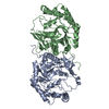
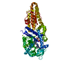
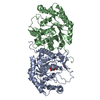
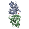
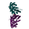
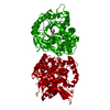
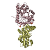
| Title | Crystal Structure of Streptococcus suis mannonate dehydratase complexed with substrate analogue | ||||||
 Components Components | D-mannonate dehydratase | ||||||
 Keywords Keywords | LYASE / Xylose isomerase-like TIM barrel | ||||||
| Function / homology |  Function and homology information Function and homology informationD-glucuronate catabolic process / mannonate dehydratase / mannonate dehydratase activity / ferrous iron binding / manganese ion binding Similarity search - Function | ||||||
| Biological species |  Streptococcus suis (bacteria) Streptococcus suis (bacteria) | ||||||
| Method |  X-RAY DIFFRACTION / MOLECULAR REPLACEMENT / Resolution: 2.5 Å X-RAY DIFFRACTION / MOLECULAR REPLACEMENT / Resolution: 2.5 Å | ||||||
 Authors Authors | Gao, F. / Zhang, Q.M. / Peng, H. / Liu, Y.W. / Qi, J.X. / Gao, G.F. | ||||||
 Citation Citation | Journal: To be Published Title: Crystal structure of Streptococcus suis mannonate dehydratase complexed with substrate analogue Authors: Gao, F. / Zhang, Q.M. / Peng, H. / Liu, Y.W. / Qi, J.X. / Gao, G.F. | ||||||
| History |
|
- Structure visualization
| Structure viewer | Molecule: MolmilJmol/JSmol |
|---|
- Downloads & links
-Download
| PDBx/mmCIF format | 3bdk.cif.gz | 147.2 KB | Display | PDBx/mmCIF format |
|---|---|---|---|---|
| PDB format | pdb3bdk.ent.gz | 115.4 KB | Display | PDB format |
| PDBx/mmJSON format | 3bdk.json.gz | Tree view | PDBx/mmJSON format | |
| Others |  Other downloads Other downloads |
-Validation report
| Summary document | 3bdk_validation.pdf.gz | 463.6 KB | Display | wwPDB validaton report |
|---|---|---|---|---|
| Full document | 3bdk_full_validation.pdf.gz | 483.5 KB | Display | |
| Data in XML | 3bdk_validation.xml.gz | 27.9 KB | Display | |
| Data in CIF | 3bdk_validation.cif.gz | 38.1 KB | Display | |
| Arichive directory | https://data.pdbj.org/pub/pdb/validation_reports/bd/3bdkftp://data.pdbj.org/pub/pdb/validation_reports/bd/3bdk | HTTPS FTP |
-Related structure data
| Related structure data | 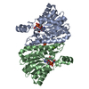 1vl8S S: Starting model for refinement |
|---|---|
| Similar structure data |


-Links
 PDBj
PDBj
- Assembly
| Deposited unit | 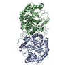
| ||||||||
|---|---|---|---|---|---|---|---|---|---|
| 1 |
| ||||||||
| Unit cell |
|
-Components
| #1: Protein | Mass: 43024.559 Da / Num. of mol.: 2 Source method: isolated from a genetically manipulated source Source: (gene. exp.) Streptococcus suis (bacteria) / Strain: strain 05ZYH33 / Gene: UxuA / Plasmid: pET28b / Production host: #2: Sugar | ChemComp-DNO / |   Type: D-saccharide / Mass: 180.156 Da / Num. of mol.: 1 Type: D-saccharide / Mass: 180.156 Da / Num. of mol.: 1Source method: isolated from a genetically manipulated source Formula: C6H12O6 #3: Chemical | ChemComp-MN / |   Mass: 54.938 Da / Num. of mol.: 1 / Source method: obtained synthetically / Formula: Mn Mass: 54.938 Da / Num. of mol.: 1 / Source method: obtained synthetically / Formula: Mn#4: Water | ChemComp-HOH / |  Mass: 18.015 Da / Num. of mol.: 41 / Source method: isolated from a natural source / Formula: H2O Mass: 18.015 Da / Num. of mol.: 41 / Source method: isolated from a natural source / Formula: H2O |
|---|
-Experimental details
-Experiment
| Experiment | Method: X-RAY DIFFRACTION |
|---|
- Sample preparation
| Crystal | Density Matthews: 2.59 Å3/Da / Density % sol: 52.47 % |
|---|---|
| Crystal grow | Method: evaporation / Details: EVAPORATION |
-Data collection
| Diffraction | Mean temperature: 100 K |
|---|---|
| Diffraction source | Source: ROTATING ANODE / Type: RIGAKU MICROMAX-007 / Wavelength: 1.5418 Å |
| Detector | Type: RIGAKU RAXIS IV++ / Detector: IMAGE PLATE |
| Radiation | Protocol: SINGLE WAVELENGTH / Monochromatic (M) / Laue (L): M / Scattering type: x-ray |
| Radiation wavelength | Wavelength: 1.5418 Å / Relative weight: 1 |
| Reflection | Resolution: 2.5→50 Å / Num. obs: 31751 / Redundancy: 13.3 % / Rmerge(I) obs: 0.063 / Net I/σ(I): 39.9 |
| Reflection shell | Resolution: 2.5→2.59 Å / Redundancy: 8.8 % / Rmerge(I) obs: 0.509 / Mean I/σ(I) obs: 2.98 / Num. unique all: 2817 |
- Processing
| Software |
| ||||||||||||||||||||||||||||||||||||||||||||||||||||||||||||||||||||||||||||||||||||||||||
|---|---|---|---|---|---|---|---|---|---|---|---|---|---|---|---|---|---|---|---|---|---|---|---|---|---|---|---|---|---|---|---|---|---|---|---|---|---|---|---|---|---|---|---|---|---|---|---|---|---|---|---|---|---|---|---|---|---|---|---|---|---|---|---|---|---|---|---|---|---|---|---|---|---|---|---|---|---|---|---|---|---|---|---|---|---|---|---|---|---|---|---|
| Refinement | Method to determine structure: MOLECULAR REPLACEMENT Starting model: 1vl8 Resolution: 2.5→45.27 Å / Cor.coef. Fo:Fc: 0.909 / Cor.coef. Fo:Fc free: 0.872 / SU B: 12.216 / SU ML: 0.276 / Cross valid method: THROUGHOUT / ESU R: 0.59 / ESU R Free: 0.344 / Stereochemistry target values: MAXIMUM LIKELIHOOD / Details: HYDROGENS HAVE BEEN ADDED IN THE RIDING POSITIONS
| ||||||||||||||||||||||||||||||||||||||||||||||||||||||||||||||||||||||||||||||||||||||||||
| Solvent computation | Ion probe radii: 0.8 Å / Shrinkage radii: 0.8 Å / VDW probe radii: 1.4 Å / Solvent model: MASK | ||||||||||||||||||||||||||||||||||||||||||||||||||||||||||||||||||||||||||||||||||||||||||
| Displacement parameters | Biso mean: 44.905 Å2
| ||||||||||||||||||||||||||||||||||||||||||||||||||||||||||||||||||||||||||||||||||||||||||
| Refinement step | Cycle: LAST / Resolution: 2.5→45.27 Å
| ||||||||||||||||||||||||||||||||||||||||||||||||||||||||||||||||||||||||||||||||||||||||||
| Refine LS restraints |
| ||||||||||||||||||||||||||||||||||||||||||||||||||||||||||||||||||||||||||||||||||||||||||
| LS refinement shell | Resolution: 2.503→2.568 Å / Total num. of bins used: 20
|
