 Movie
Movie Controller

[English] 日本語
 Yorodumi
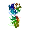
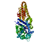
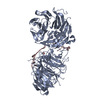
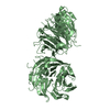
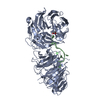
- PDB-3ban: The crystal structure of mannonate dehydratase from Streptococcus... -
+ Open data

- Basic information
| Entry | Database: PDB / ID: 3ban | ||||||
|---|---|---|---|---|---|---|---|
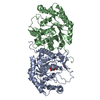
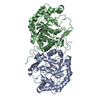
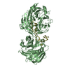
| Title | The crystal structure of mannonate dehydratase from Streptococcus suis serotype2 | ||||||
 Components Components | D-mannonate dehydratase | ||||||
 Keywords Keywords | LYASE / xylose-like TIM barrel | ||||||
| Function / homology |  Function and homology information Function and homology informationD-glucuronate catabolic process / mannonate dehydratase / mannonate dehydratase activity / ferrous iron binding / manganese ion binding Similarity search - Function | ||||||
| Biological species |  Streptococcus suis (bacteria) Streptococcus suis (bacteria) | ||||||
| Method |  X-RAY DIFFRACTION / MOLECULAR REPLACEMENT / Resolution: 2.9 Å X-RAY DIFFRACTION / MOLECULAR REPLACEMENT / Resolution: 2.9 Å | ||||||
 Authors Authors | Peng, H. / Zhang, Q.M. / Gao, F. / Liu, Y.W. / Qi, J.X. / Gao, G.F. | ||||||
 Citation Citation | Journal: To be Published Title: The crystal structure of mannonate dehydratase from Streptococcus suis serotype2 Authors: Peng, H. / Zhang, Q.M. / Gao, F. / Liu, Y.W. / Qi, J.X. / Gao, G.F. | ||||||
| History |
|
- Structure visualization
| Structure viewer | Molecule: MolmilJmol/JSmol |
|---|
- Downloads & links
-Download
| PDBx/mmCIF format | 3ban.cif.gz | 144.1 KB | Display | PDBx/mmCIF format |
|---|---|---|---|---|
| PDB format | pdb3ban.ent.gz | 114.5 KB | Display | PDB format |
| PDBx/mmJSON format | 3ban.json.gz | Tree view | PDBx/mmJSON format | |
| Others |  Other downloads Other downloads |
-Validation report
| Arichive directory | https://data.pdbj.org/pub/pdb/validation_reports/ba/3banftp://data.pdbj.org/pub/pdb/validation_reports/ba/3ban | HTTPS FTP |
|---|
-Related structure data
| Related structure data | 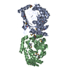 1tz9S S: Starting model for refinement |
|---|---|
| Similar structure data |


-Links
 PDBj
PDBj
- Assembly
| Deposited unit | 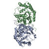
| ||||||||
|---|---|---|---|---|---|---|---|---|---|
| 1 |
| ||||||||
| Unit cell |
|
-Components
| #1: Protein | Mass: 43024.559 Da / Num. of mol.: 2 Source method: isolated from a genetically manipulated source Source: (gene. exp.) Streptococcus suis (bacteria) / Strain: Serotype 2, strain 05ZYH33 / Gene: UxuA / Plasmid: pET28b / Production host: |
|---|
-Experimental details
-Experiment
| Experiment | Method: X-RAY DIFFRACTION |
|---|
- Sample preparation
| Crystal | Density Matthews: 2.59 Å3/Da / Density % sol: 52.5 % |
|---|
-Data collection
| Diffraction source | Source: ROTATING ANODE / Type: RIGAKU MICROMAX-007 |
|---|---|
| Radiation | Protocol: SINGLE WAVELENGTH / Monochromatic (M) / Laue (L): M / Scattering type: x-ray |
| Radiation wavelength | Relative weight: 1 |
| Reflection | Resolution: 2.9→105.409 Å / Num. obs: 20728 / Redundancy: 13.85 % / Biso Wilson estimate: 61.2 Å2 / Rmerge(I) obs: 0.12 / Net I/σ(I): 11.8 |
| Reflection shell | Resolution: 2.9→3 Å / Redundancy: 14.13 % / Rmerge(I) obs: 0.37 / Mean I/σ(I) obs: 5.7 |
- Processing
| Software |
| ||||||||||||||||||||||||||||||||||||||||||||||||||||||||||||||||||||||||||||||||||||||||||
|---|---|---|---|---|---|---|---|---|---|---|---|---|---|---|---|---|---|---|---|---|---|---|---|---|---|---|---|---|---|---|---|---|---|---|---|---|---|---|---|---|---|---|---|---|---|---|---|---|---|---|---|---|---|---|---|---|---|---|---|---|---|---|---|---|---|---|---|---|---|---|---|---|---|---|---|---|---|---|---|---|---|---|---|---|---|---|---|---|---|---|---|
| Refinement | Method to determine structure: MOLECULAR REPLACEMENT Starting model: PDB ENTRY 1TZ9 Resolution: 2.9→44.06 Å / Cor.coef. Fo:Fc: 0.888 / Cor.coef. Fo:Fc free: 0.854 / SU B: 18.594 / SU ML: 0.361 / Cross valid method: THROUGHOUT / ESU R Free: 0.446 / Stereochemistry target values: MAXIMUM LIKELIHOOD / Details: HYDROGENS HAVE BEEN ADDED IN THE RIDING POSITIONS
| ||||||||||||||||||||||||||||||||||||||||||||||||||||||||||||||||||||||||||||||||||||||||||
| Solvent computation | Ion probe radii: 0.8 Å / Shrinkage radii: 0.8 Å / VDW probe radii: 1.4 Å / Solvent model: MASK | ||||||||||||||||||||||||||||||||||||||||||||||||||||||||||||||||||||||||||||||||||||||||||
| Displacement parameters | Biso mean: 41.716 Å2
| ||||||||||||||||||||||||||||||||||||||||||||||||||||||||||||||||||||||||||||||||||||||||||
| Refinement step | Cycle: LAST / Resolution: 2.9→44.06 Å
| ||||||||||||||||||||||||||||||||||||||||||||||||||||||||||||||||||||||||||||||||||||||||||
| Refine LS restraints |
| ||||||||||||||||||||||||||||||||||||||||||||||||||||||||||||||||||||||||||||||||||||||||||
| LS refinement shell | Resolution: 2.9→2.975 Å / Total num. of bins used: 20
|
