oxidoreductase activity, acting on NAD(P)H, oxygen as acceptor / oxidoreductase activity, acting on the CH-OH group of donors, NAD or NADP as acceptor / carbohydrate metabolic process / metal ion binding Similarity search - Function
Resolution: 1.8→1.83 Å / Redundancy: 6.9 % / Rmerge(I) obs: 0.503 / Mean I/σ(I) obs: 4.03 / Num. unique all: 4828 / % possible all: 99.9
-
Processing
Software
Name
Version
Classification
HKL-2000
datacollection
PHASES
phasing
REFMAC
5.2.0019
refinement
HKL-2000
datareduction
HKL-2000
datascaling
Refinement
Method to determine structure: MOLECULAR REPLACEMENT / Resolution: 1.8→50 Å / Cor.coef. Fo:Fc: 0.965 / Cor.coef. Fo:Fc free: 0.947 / SU B: 3.875 / SU ML: 0.064 / Cross valid method: THROUGHOUT / ESU R Free: 0.104 / Stereochemistry target values: MAXIMUM LIKELIHOOD / Details: HYDROGENS HAVE BEEN ADDED IN THE RIDING POSITIONS
Rfactor
Num. reflection
% reflection
Selection details
Rfree
0.18448
4874
5 %
RANDOM
Rwork
0.15053
-
-
-
all
0.15224
92750
-
-
obs
0.15224
92746
99.64 %
-
Solvent computation
Ion probe radii: 0.8 Å / Shrinkage radii: 0.8 Å / VDW probe radii: 1.2 Å / Solvent model: MASK
Displacement parameters
Biso mean: 18.204 Å2
Baniso -1
Baniso -2
Baniso -3
1-
-0.74 Å2
0 Å2
0 Å2
2-
-
1.35 Å2
0 Å2
3-
-
-
-0.61 Å2
Refinement step
Cycle: LAST / Resolution: 1.8→50 Å
Protein
Nucleic acid
Ligand
Solvent
Total
Num. atoms
7704
0
4
1084
8792
Refine LS restraints
Refine-ID
Type
Dev ideal
Dev ideal target
Number
X-RAY DIFFRACTION
r_bond_refined_d
0.012
0.022
7808
X-RAY DIFFRACTION
r_angle_refined_deg
1.189
1.943
10596
X-RAY DIFFRACTION
r_dihedral_angle_1_deg
5.681
5
1024
X-RAY DIFFRACTION
r_dihedral_angle_2_deg
40.552
24.881
336
X-RAY DIFFRACTION
r_dihedral_angle_3_deg
12.26
15
1316
X-RAY DIFFRACTION
r_dihedral_angle_4_deg
16.819
15
52
X-RAY DIFFRACTION
r_chiral_restr
0.081
0.2
1244
X-RAY DIFFRACTION
r_gen_planes_refined
0.005
0.02
5860
X-RAY DIFFRACTION
r_nbd_refined
0.199
0.2
4140
X-RAY DIFFRACTION
r_nbtor_refined
0.298
0.2
5495
X-RAY DIFFRACTION
r_xyhbond_nbd_refined
0.128
0.2
848
X-RAY DIFFRACTION
r_symmetry_vdw_refined
0.238
0.2
64
X-RAY DIFFRACTION
r_symmetry_hbond_refined
0.236
0.2
49
X-RAY DIFFRACTION
r_mcbond_it
0.76
1.5
5251
X-RAY DIFFRACTION
r_mcangle_it
1.1
2
8136
X-RAY DIFFRACTION
r_scbond_it
2.044
3
2937
X-RAY DIFFRACTION
r_scangle_it
3.28
4.5
2460
LS refinement shell
Resolution: 1.801→1.848 Å / Total num. of bins used: 20
Rfactor
Num. reflection
% reflection
Rfree
0.264
350
-
Rwork
0.204
6627
-
obs
-
-
97.48 %
Refinement TLS params.
Method: refined / Origin x: 3.871 Å / Origin y: 50.734 Å / Origin z: 26.4957 Å
11
12
13
21
22
23
31
32
33
T
-0.1117 Å2
-0.0162 Å2
0.0101 Å2
-
-0.0987 Å2
-0.0013 Å2
-
-
-0.0971 Å2
L
0.3513 °2
-0.1228 °2
0.1588 °2
-
0.4033 °2
-0.2352 °2
-
-
0.5157 °2
S
-0.009 Å °
0.0783 Å °
-0.0427 Å °
-0.002 Å °
-0.055 Å °
-0.0391 Å °
0.0054 Å °
0.0721 Å °
0.064 Å °
Refinement TLS group
ID
Refine-ID
Refine TLS-ID
Auth asym-ID
Auth seq-ID
1
X-RAY DIFFRACTION
1
A
1 - 257
2
X-RAY DIFFRACTION
1
B
1 - 257
3
X-RAY DIFFRACTION
1
C
1 - 257
4
X-RAY DIFFRACTION
1
D
1 - 257
+
About Yorodumi
-
News
-
Feb 9, 2022. New format data for meta-information of EMDB entries
New format data for meta-information of EMDB entries
Version 3 of the EMDB header file is now the official format.
The previous official version 1.9 will be removed from the archive.
In the structure databanks used in Yorodumi, some data are registered as the other names, "COVID-19 virus" and "2019-nCoV". Here are the details of the virus and the list of structure data.
Jan 31, 2019. EMDB accession codes are about to change! (news from PDBe EMDB page)
EMDB accession codes are about to change! (news from PDBe EMDB page)
The allocation of 4 digits for EMDB accession codes will soon come to an end. Whilst these codes will remain in use, new EMDB accession codes will include an additional digit and will expand incrementally as the available range of codes is exhausted. The current 4-digit format prefixed with “EMD-” (i.e. EMD-XXXX) will advance to a 5-digit format (i.e. EMD-XXXXX), and so on. It is currently estimated that the 4-digit codes will be depleted around Spring 2019, at which point the 5-digit format will come into force.
The EM Navigator/Yorodumi systems omit the EMD- prefix.
Related info.:Q: What is EMD? / ID/Accession-code notation in Yorodumi/EM Navigator
Yorodumi is a browser for structure data from EMDB, PDB, SASBDB, etc.
This page is also the successor to EM Navigator detail page, and also detail information page/front-end page for Omokage search.
The word "yorodu" (or yorozu) is an old Japanese word meaning "ten thousand". "mi" (miru) is to see.
Related info.:EMDB / PDB / SASBDB / Comparison of 3 databanks / Yorodumi Search / Aug 31, 2016. New EM Navigator & Yorodumi / Yorodumi Papers / Jmol/JSmol / Function and homology information / Changes in new EM Navigator and Yorodumi
 Movie
Movie Controller


 Open data
 Basic information
 Components
 Keywords
 Function and homology information
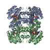
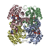
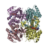
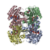
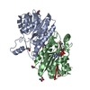
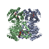
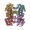
 Gluconobacter oxydans (bacteria)
 X-RAY DIFFRACTION /
  Authors
 Citation
 Structure visualization
 Downloads & links
 Other downloads
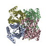

 PDBj
PDBj


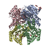
 Assembly
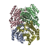

 Mass: 24.305 Da / Num. of mol.: 2 / Source method: obtained synthetically / Formula: Mg
 Mass: 112.411 Da / Num. of mol.: 2 / Source method: obtained synthetically / Formula: Cd
 Mass: 18.015 Da / Num. of mol.: 1084 / Source method: isolated from a natural source / Formula: H2O
 Sample preparation
 / Beamline: X29A / Wavelength: 1.075 Å
 Processing