 Movie
Movie Controller

[English] 日本語
 Yorodumi
- PDB-3av3: Crystal structure of glycinamide ribonucleotide transformylase 1 ... -
+ Open data

- Basic information
| Entry | Database: PDB / ID: 3av3 | ||||||
|---|---|---|---|---|---|---|---|
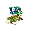
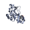
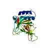
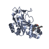
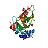
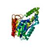
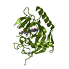
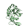
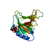
| Title | Crystal structure of glycinamide ribonucleotide transformylase 1 from Geobacillus kaustophilus | ||||||
 Components Components | Phosphoribosylglycinamide formyltransferase | ||||||
 Keywords Keywords | TRANSFERASE / Structural Genomics / RIKEN Structural Genomics/Proteomics Initiative / RSGI / Rossmann fold / transformylase / folate binding | ||||||
| Function / homology |  Function and homology informationphosphoribosylglycinamide formyltransferase 1 / phosphoribosylglycinamide formyltransferase activity / 'de novo' IMP biosynthetic process Function and homology informationphosphoribosylglycinamide formyltransferase 1 / phosphoribosylglycinamide formyltransferase activity / 'de novo' IMP biosynthetic processSimilarity search - Function | ||||||
| Biological species |  Geobacillus kaustophilus (bacteria) Geobacillus kaustophilus (bacteria) | ||||||
| Method | X-RAY DIFFRACTION / SYNCHROTRON / MAD / Resolution: 1.7 Å | ||||||
 Authors Authors | Kanagawa, M. / Baba, S. / Nakagawa, N. / Ebihara, A. / Kuramitsu, S. / Yokoyama, S. / Sampei, G. / Kawai, G. / RIKEN Structural Genomics/Proteomics Initiative (RSGI) | ||||||
 Citation Citation | Journal: J.Biochem. / Year: 2013 Title: Structures and reaction mechanisms of the two related enzymes, PurN and PurU. Authors: Sampei, G. / Kanagawa, M. / Baba, S. / Shimasaki, T. / Taka, H. / Mitsui, S. / Fujiwara, S. / Yanagida, Y. / Kusano, M. / Suzuki, S. / Terao, K. / Kawai, H. / Fukai, Y. / Nakagawa, N. / ...Authors: Sampei, G. / Kanagawa, M. / Baba, S. / Shimasaki, T. / Taka, H. / Mitsui, S. / Fujiwara, S. / Yanagida, Y. / Kusano, M. / Suzuki, S. / Terao, K. / Kawai, H. / Fukai, Y. / Nakagawa, N. / Ebihara, A. / Kuramitsu, S. / Yokoyama, S. / Kawai, G. | ||||||
| History |
|
- Structure visualization
| Structure viewer | Molecule: MolmilJmol/JSmol |
|---|
- Downloads & links
-Download
| PDBx/mmCIF format | 3av3.cif.gz | 53.4 KB | Display | PDBx/mmCIF format |
|---|---|---|---|---|
| PDB format | pdb3av3.ent.gz | 41.1 KB | Display | PDB format |
| PDBx/mmJSON format | 3av3.json.gz | Tree view | PDBx/mmJSON format | |
| Others |  Other downloads Other downloads |
-Validation report
| Arichive directory | https://data.pdbj.org/pub/pdb/validation_reports/av/3av3ftp://data.pdbj.org/pub/pdb/validation_reports/av/3av3 | HTTPS FTP |
|---|
-Related structure data


-Links
 PDBj
PDBj- Assembly
| Deposited unit | 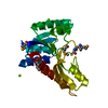
| |||||||||||||||
|---|---|---|---|---|---|---|---|---|---|---|---|---|---|---|---|---|
| 1 |
| |||||||||||||||
| Unit cell |
| |||||||||||||||
| Components on special symmetry positions |
|
-Components
| #1: Protein | Mass: 23469.111 Da / Num. of mol.: 1 Source method: isolated from a genetically manipulated source Source: (gene. exp.) Geobacillus kaustophilus (bacteria) / Strain: HTA426 / Gene: GK0266 / Plasmid: pET-HisTEV / Production host: Escherichia coli (E. coli)References: UniProt: Q5L3C9, phosphoribosylglycinamide formyltransferase 1 | ||
|---|---|---|---|
| #2: Chemical |   Mass: 24.305 Da / Num. of mol.: 2 / Source method: obtained synthetically / Formula: Mg Mass: 24.305 Da / Num. of mol.: 2 / Source method: obtained synthetically / Formula: Mg#3: Water | ChemComp-HOH / | Water Mass: 18.015 Da / Num. of mol.: 275 / Source method: isolated from a natural source / Formula: H2O Mass: 18.015 Da / Num. of mol.: 275 / Source method: isolated from a natural source / Formula: H2O |
-Experimental details
-Experiment
| Experiment | Method: X-RAY DIFFRACTION / Number of used crystals: 1 |
|---|
- Sample preparation
| Crystal | Density Matthews: 2.18 Å3/Da / Density % sol: 43.6 % / Description: The file contains Friedel pairs. |
|---|---|
| Crystal grow | Method: vapor diffusion, sitting drop / pH: 6.4 Details: Bis-Tris 0.1M pH 6.4, 0.5M Sodium Fluoride, PEG 3350 25%, Mg Chloride 0.1M, VAPOR DIFFUSION, SITTING DROP |
-Data collection
| Diffraction | Mean temperature: 100 K | ||||||||||||
|---|---|---|---|---|---|---|---|---|---|---|---|---|---|
| Diffraction source | Source: SYNCHROTRON / Site: SPring-8  / Beamline: BL26B1 / Wavelength: 0.97944, 0.90000, 0.97984 / Beamline: BL26B1 / Wavelength: 0.97944, 0.90000, 0.97984 | ||||||||||||
| Detector | Type: RIGAKU JUPITER 210 / Detector: CCD / Date: Feb 15, 2008 / Details: Toroidal Mirror | ||||||||||||
| Radiation | Monochromator: Fixed exit Si 111 double crystal monochromater Protocol: MAD / Monochromatic (M) / Laue (L): M / Scattering type: x-ray | ||||||||||||
| Radiation wavelength |
| ||||||||||||
| Reflection | Resolution: 1.7→50 Å / Num. obs: 43718 / % possible obs: 99.7 % / Redundancy: 7 % / Biso Wilson estimate: 17.2 Å2 / Rmerge(I) obs: 0.045 | ||||||||||||
| Reflection shell | Resolution: 1.7→1.76 Å / Redundancy: 6.6 % / Rmerge(I) obs: 0.115 / Mean I/σ(I) obs: 14.7 / % possible all: 98.6 |
- Processing
| Software |
| ||||||||||||||||||||||||||||||||||||||||||||||||||||||||||||
|---|---|---|---|---|---|---|---|---|---|---|---|---|---|---|---|---|---|---|---|---|---|---|---|---|---|---|---|---|---|---|---|---|---|---|---|---|---|---|---|---|---|---|---|---|---|---|---|---|---|---|---|---|---|---|---|---|---|---|---|---|---|
| Refinement | Method to determine structure: MAD / Resolution: 1.7→38.77 Å / Rfactor Rfree error: 0.003 / Data cutoff high absF: 237504.67 / Data cutoff low absF: 0 / Isotropic thermal model: RESTRAINED / Cross valid method: THROUGHOUT / σ(F): 0 Details: The file contains Friedel pairs. BULK SOLVENT MODEL USED.
| ||||||||||||||||||||||||||||||||||||||||||||||||||||||||||||
| Solvent computation | Solvent model: FLAT MODEL / Bsol: 55.4148 Å2 / ksol: 0.4 e/Å3 | ||||||||||||||||||||||||||||||||||||||||||||||||||||||||||||
| Displacement parameters | Biso mean: 19.5 Å2
| ||||||||||||||||||||||||||||||||||||||||||||||||||||||||||||
| Refine analyze |
| ||||||||||||||||||||||||||||||||||||||||||||||||||||||||||||
| Refinement step | Cycle: LAST / Resolution: 1.7→38.77 Å
| ||||||||||||||||||||||||||||||||||||||||||||||||||||||||||||
| Refine LS restraints |
| ||||||||||||||||||||||||||||||||||||||||||||||||||||||||||||
| LS refinement shell | Resolution: 1.7→1.81 Å / Rfactor Rfree error: 0.01 / Total num. of bins used: 6
| ||||||||||||||||||||||||||||||||||||||||||||||||||||||||||||
| Xplor file |
|
