 Movie
Movie Controller

[English] 日本語
 Yorodumi
- PDB-3a38: Crystal structure of high-potential iron-sulfur protein from Ther... -
+ Open data

- Basic information
| Entry | Database: PDB / ID: 3a38 | ||||||
|---|---|---|---|---|---|---|---|
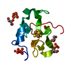
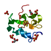
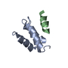
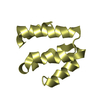
| Title | Crystal structure of high-potential iron-sulfur protein from Thermochromatium tepidum at 0.7 angstrom resolution | ||||||
 Components Components | High-potential iron-sulfur protein | ||||||
 Keywords Keywords | ELECTRON TRANSPORT / IRON-SULFUR CLUSTER / Iron / Iron-sulfur / Metal-binding / Transport | ||||||
| Function / homology |  Function and homology information Function and homology informationaerobic electron transport chain / 4 iron, 4 sulfur cluster binding / electron transfer activity / metal ion binding Similarity search - Function | ||||||
| Biological species |  Thermochromatium tepidum (bacteria) Thermochromatium tepidum (bacteria) | ||||||
| Method |  X-RAY DIFFRACTION / SYNCHROTRON / MOLECULAR REPLACEMENT / Resolution: 0.7 Å X-RAY DIFFRACTION / SYNCHROTRON / MOLECULAR REPLACEMENT / Resolution: 0.7 Å | ||||||
 Authors Authors | Takeda, K. / Kusumoto, K. / Hirano, Y. / Miki, K. | ||||||
 Citation Citation | Journal: J.Struct.Biol. / Year: 2010 Title: Detailed assessment of X-ray induced structural perturbation in a crystalline state protein. Authors: Takeda, K. / Kusumoto, K. / Hirano, Y. / Miki, K. | ||||||
| History |
|
- Structure visualization
| Structure viewer | Molecule: MolmilJmol/JSmol |
|---|
- Downloads & links
-Download
| PDBx/mmCIF format | 3a38.cif.gz | 71.3 KB | Display | PDBx/mmCIF format |
|---|---|---|---|---|
| PDB format | pdb3a38.ent.gz | 52.8 KB | Display | PDB format |
| PDBx/mmJSON format | 3a38.json.gz | Tree view | PDBx/mmJSON format | |
| Others |  Other downloads Other downloads |
-Validation report
| Summary document | 3a38_validation.pdf.gz | 456.9 KB | Display | wwPDB validaton report |
|---|---|---|---|---|
| Full document | 3a38_full_validation.pdf.gz | 458.7 KB | Display | |
| Data in XML | 3a38_validation.xml.gz | 8.6 KB | Display | |
| Data in CIF | 3a38_validation.cif.gz | 11.9 KB | Display | |
| Arichive directory | https://data.pdbj.org/pub/pdb/validation_reports/a3/3a38ftp://data.pdbj.org/pub/pdb/validation_reports/a3/3a38 | HTTPS FTP |
-Related structure data
| Related structure data |  3a39C  1iuaS C: citing same article ( S: Starting model for refinement |
|---|---|
| Similar structure data |


-Links
 PDBj
PDBj- Assembly
| Deposited unit | 
| ||||||||
|---|---|---|---|---|---|---|---|---|---|
| 1 |
| ||||||||
| Unit cell |
|
-Components
| #1: Protein | Mass: 8793.851 Da / Num. of mol.: 1 / Source method: isolated from a natural source / Source: (natural) Thermochromatium tepidum (bacteria) / References: UniProt: P80176 | ||||
|---|---|---|---|---|---|
| #2: Chemical | ChemComp-SF4 /   Mass: 351.640 Da / Num. of mol.: 1 / Source method: obtained synthetically / Formula: Fe4S4 Mass: 351.640 Da / Num. of mol.: 1 / Source method: obtained synthetically / Formula: Fe4S4 | ||||
| #3: Chemical |   Mass: 96.063 Da / Num. of mol.: 3 / Source method: obtained synthetically / Formula: SO4 Mass: 96.063 Da / Num. of mol.: 3 / Source method: obtained synthetically / Formula: SO4#4: Chemical | ChemComp-GOL / |   Mass: 92.094 Da / Num. of mol.: 1 / Source method: obtained synthetically / Formula: C3H8O3 Mass: 92.094 Da / Num. of mol.: 1 / Source method: obtained synthetically / Formula: C3H8O3#5: Water | ChemComp-HOH / |  Mass: 18.015 Da / Num. of mol.: 162 / Source method: isolated from a natural source / Formula: H2O Mass: 18.015 Da / Num. of mol.: 162 / Source method: isolated from a natural source / Formula: H2O |
-Experimental details
-Experiment
| Experiment | Method: X-RAY DIFFRACTION / Number of used crystals: 1 |
|---|
- Sample preparation
| Crystal | Density Matthews: 1.51 Å3/Da / Density % sol: 32.61 % |
|---|---|
| Crystal grow | Temperature: 293 K / Method: vapor diffusion, hanging drop / pH: 4 Details: 1.9M ammonium sulfate, 10mM dithiothreitol, 100mM sodium citrate, pH 4.0, VAPOR DIFFUSION, HANGING DROP, temperature 293K |
-Data collection
| Diffraction | Mean temperature: 93 K |
|---|---|
| Diffraction source | Source: SYNCHROTRON / Site: SPring-8  / Beamline: BL41XU / Wavelength: 0.71 Å / Beamline: BL41XU / Wavelength: 0.71 Å |
| Detector | Type: ADSC QUANTUM 315 / Detector: CCD |
| Radiation | Protocol: SINGLE WAVELENGTH / Monochromatic (M) / Laue (L): M / Scattering type: x-ray |
| Radiation wavelength | Wavelength: 0.71 Å / Relative weight: 1 |
| Reflection | Resolution: 0.7→50 Å / Num. obs: 96778 / % possible obs: 94.7 % / Redundancy: 6.2 % / Rmerge(I) obs: 0.055 / Net I/σ(I): 57.2 |
| Reflection shell | Resolution: 0.7→0.73 Å / Redundancy: 1.9 % / Rmerge(I) obs: 0.156 / Mean I/σ(I) obs: 8.9 / Num. unique all: 6229 / % possible all: 61.7 |
- Processing
| Software |
| |||||||||||||||||||||||||||||||||
|---|---|---|---|---|---|---|---|---|---|---|---|---|---|---|---|---|---|---|---|---|---|---|---|---|---|---|---|---|---|---|---|---|---|---|
| Refinement | Method to determine structure: MOLECULAR REPLACEMENT Starting model: 1IUA Resolution: 0.7→50 Å / Num. parameters: 8703 / Num. restraintsaints: 413 / Cross valid method: THROUGHOUT / σ(F): 0 / Stereochemistry target values: Engh & Huber
| |||||||||||||||||||||||||||||||||
| Refine analyze | Num. disordered residues: 38 / Occupancy sum hydrogen: 544.02 / Occupancy sum non hydrogen: 744.38 | |||||||||||||||||||||||||||||||||
| Refinement step | Cycle: LAST / Resolution: 0.7→50 Å
| |||||||||||||||||||||||||||||||||
| Refine LS restraints |
|
