 Movie
Movie Controller

[English] 日本語
 Yorodumi
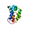
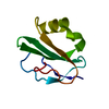
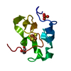
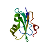
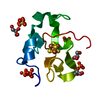
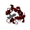
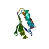
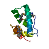
- PDB-1eyt: CRYSTAL STRUCTURE OF HIGH-POTENTIAL IRON-SULFUR PROTEIN FROM THER... -
+ Open data

- Basic information
| Entry | Database: PDB / ID: 1eyt | ||||||
|---|---|---|---|---|---|---|---|
| Title | CRYSTAL STRUCTURE OF HIGH-POTENTIAL IRON-SULFUR PROTEIN FROM THERMOCHROMATIUM TEPIDUM | ||||||
 Components Components | HIGH-POTENTIAL IRON-SULFUR PROTEIN | ||||||
 Keywords Keywords | ELECTRON TRANSPORT / Iron-sulfur protein | ||||||
| Function / homology |  Function and homology information Function and homology informationaerobic electron transport chain / 4 iron, 4 sulfur cluster binding / electron transfer activity / metal ion binding Similarity search - Function | ||||||
| Biological species |  Thermochromatium tepidum (bacteria) Thermochromatium tepidum (bacteria) | ||||||
| Method |  X-RAY DIFFRACTION / SYNCHROTRON / Resolution: 1.5 Å X-RAY DIFFRACTION / SYNCHROTRON / Resolution: 1.5 Å | ||||||
 Authors Authors | Nogi, T. / Fathir, I. / Kobayashi, M. / Nozawa, T. / Miki, K. | ||||||
 Citation Citation | Journal: Proc.Natl.Acad.Sci.USA / Year: 2000 Title: Crystal structures of photosynthetic reaction center and high-potential iron-sulfur protein from Thermochromatium tepidum: thermostability and electron transfer. Authors: Nogi, T. / Fathir, I. / Kobayashi, M. / Nozawa, T. / Miki, K. #1: Journal: Acta Crystallogr.,Sect.D / Year: 2000Title: Crystallization and Preliminary Crystallographic Analysis of High-potential Iron-sulfur Protein from Thermochromatium tepidum Authors: Nogi, T. / Kobayashi, M. / Nozawa, T. / Miki, K. | ||||||
| History |
|
- Structure visualization
| Structure viewer | Molecule: MolmilJmol/JSmol |
|---|
- Downloads & links
-Download
| PDBx/mmCIF format | 1eyt.cif.gz | 28.1 KB | Display | PDBx/mmCIF format |
|---|---|---|---|---|
| PDB format | pdb1eyt.ent.gz | 17.3 KB | Display | PDB format |
| PDBx/mmJSON format | 1eyt.json.gz | Tree view | PDBx/mmJSON format | |
| Others |  Other downloads Other downloads |
-Validation report
| Arichive directory | https://data.pdbj.org/pub/pdb/validation_reports/ey/1eytftp://data.pdbj.org/pub/pdb/validation_reports/ey/1eyt | HTTPS FTP |
|---|
-Related structure data


-Links
 PDBj
PDBj- Assembly
| Deposited unit | 
| ||||||||
|---|---|---|---|---|---|---|---|---|---|
| 1 |
| ||||||||
| Unit cell |
|
-Components
| #1: Protein | Mass: 8793.851 Da / Num. of mol.: 1 / Source method: isolated from a natural source / Source: (natural) Thermochromatium tepidum (bacteria) / References: UniProt: P80176 |
|---|---|
| #2: Chemical | ChemComp-SF4 /   Mass: 351.640 Da / Num. of mol.: 1 / Source method: obtained synthetically / Formula: Fe4S4 Mass: 351.640 Da / Num. of mol.: 1 / Source method: obtained synthetically / Formula: Fe4S4 |
| #3: Water | ChemComp-HOH /  Mass: 18.015 Da / Num. of mol.: 43 / Source method: isolated from a natural source / Formula: H2O Mass: 18.015 Da / Num. of mol.: 43 / Source method: isolated from a natural source / Formula: H2O |
-Experimental details
-Experiment
| Experiment | Method: X-RAY DIFFRACTION / Number of used crystals: 1 |
|---|
- Sample preparation
| Crystal | Density Matthews: 1.88 Å3/Da / Density % sol: 34.72 % | ||||||||||||||||||||||||||||||
|---|---|---|---|---|---|---|---|---|---|---|---|---|---|---|---|---|---|---|---|---|---|---|---|---|---|---|---|---|---|---|---|
| Crystal grow | Temperature: 293 K / Method: vapor diffusion, sitting drop / pH: 3.5 Details: ammonium sulfate, sodium citrate, pH 3.5, VAPOR DIFFUSION, SITTING DROP, temperature 293.0K | ||||||||||||||||||||||||||||||
| Crystal grow | *PLUS Temperature: 297 K / Method: vapor diffusionDetails: Nogi, T., (2000) Acta Crystallogr., Sect.D, 56, 656. | ||||||||||||||||||||||||||||||
| Components of the solutions | *PLUS
|
-Data collection
| Diffraction | Mean temperature: 293 K |
|---|---|
| Diffraction source | Source: SYNCHROTRON / Site: Photon Factory  / Beamline: BL-6A / Wavelength: 1 / Beamline: BL-6A / Wavelength: 1 |
| Detector | Type: WEISSENBERG / Detector: DIFFRACTOMETER / Date: Nov 17, 1999 |
| Radiation | Protocol: SINGLE WAVELENGTH / Monochromatic (M) / Laue (L): M / Scattering type: x-ray |
| Radiation wavelength | Wavelength: 1 Å / Relative weight: 1 |
| Reflection | Resolution: 1.5→20 Å / Num. all: 10306 / Num. obs: 9746 / % possible obs: 86.7 % / Observed criterion σ(F): 2 / Observed criterion σ(I): 1 / Redundancy: 4.6 % / Biso Wilson estimate: 8.4 Å2 / Rmerge(I) obs: 0.067 / Net I/σ(I): 13.1 |
| Reflection shell | Resolution: 1.5→1.53 Å / Redundancy: 2.8 % / Rmerge(I) obs: 0.219 / Num. unique all: 561 / % possible all: 67.6 |
| Reflection | *PLUS Num. obs: 10306 / Num. measured all: 50767 |
| Reflection shell | *PLUS % possible obs: 67.6 % / Mean I/σ(I) obs: 6 |
- Processing
| Software |
| ||||||||||||||||||||
|---|---|---|---|---|---|---|---|---|---|---|---|---|---|---|---|---|---|---|---|---|---|
| Refinement | Resolution: 1.5→10 Å / σ(F): 2 / σ(I): 1 / Stereochemistry target values: Engh & Huber
| ||||||||||||||||||||
| Refinement step | Cycle: LAST / Resolution: 1.5→10 Å
| ||||||||||||||||||||
| Refine LS restraints |
| ||||||||||||||||||||
| Software | *PLUS Name: X-PLOR / Version: 3.851 / Classification: refinement | ||||||||||||||||||||
| Refine LS restraints | *PLUS
| ||||||||||||||||||||
| LS refinement shell | *PLUS Highest resolution: 1.5 Å / Lowest resolution: 1.57 Å / Rfactor Rfree: 0.248 / Rfactor Rwork: 0.247 |
