 Movie
Movie Controller

[English] 日本語
 Yorodumi
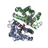
- PDB-2z8a: Ligand Migration and Binding in The Dimeric Hemoglobin of Scaphar... -
+ Open data

- Basic information
| Entry | Database: PDB / ID: 2z8a | ||||||
|---|---|---|---|---|---|---|---|
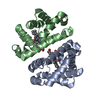
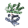
| Title | Ligand Migration and Binding in The Dimeric Hemoglobin of Scapharca Inaequivalvis: I25W with CO Bound to HEME and in the Presence of 3 Atoms of XE | ||||||
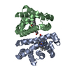
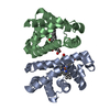
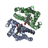
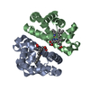
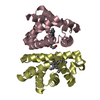
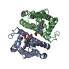
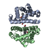
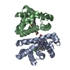
 Components Components | Globin-1 | ||||||
 Keywords Keywords | OXYGEN BINDING / OXYGEN TRANSPORT / ALLOSTERY / OXYGEN AFFINITY / Cytoplasm / Heme / Iron / Metal-binding / OXYGEN STORAGE/TRANSPORT | ||||||
| Function / homology |  Function and homology information Function and homology informationoxygen carrier activity / oxygen binding / response to hypoxia / heme binding / identical protein binding / metal ion binding / cytoplasm Similarity search - Function | ||||||
| Biological species |  Scapharca inaequivalvis (ark clam) Scapharca inaequivalvis (ark clam) | ||||||
| Method |  X-RAY DIFFRACTION / SYNCHROTRON / MOLECULAR REPLACEMENT / Resolution: 1.06 Å X-RAY DIFFRACTION / SYNCHROTRON / MOLECULAR REPLACEMENT / Resolution: 1.06 Å | ||||||
 Authors Authors | Knapp, J.E. / Royer Jr., W.E. / Nienhaus, K. / Palladino, P. / Nienhaus, G.U. | ||||||
 Citation Citation | Journal: Biochemistry / Year: 2007 Title: Ligand Migration and Binding in the Dimeric Hemoglobin of Scapharca inaequivalvis Authors: Nienhaus, K. / Knapp, J.E. / Palladino, P. / Royer Jr., W.E. / Nienhaus, G.U. | ||||||
| History |
|
- Structure visualization
| Structure viewer | Molecule: MolmilJmol/JSmol |
|---|
- Downloads & links
-Download
| PDBx/mmCIF format | 2z8a.cif.gz | 147.5 KB | Display | PDBx/mmCIF format |
|---|---|---|---|---|
| PDB format | pdb2z8a.ent.gz | 114 KB | Display | PDB format |
| PDBx/mmJSON format | 2z8a.json.gz | Tree view | PDBx/mmJSON format | |
| Others |  Other downloads Other downloads |
-Validation report
| Summary document | 2z8a_validation.pdf.gz | 1.1 MB | Display | wwPDB validaton report |
|---|---|---|---|---|
| Full document | 2z8a_full_validation.pdf.gz | 1.1 MB | Display | |
| Data in XML | 2z8a_validation.xml.gz | 17.6 KB | Display | |
| Data in CIF | 2z8a_validation.cif.gz | 25.7 KB | Display | |
| Arichive directory | https://data.pdbj.org/pub/pdb/validation_reports/z8/2z8aftp://data.pdbj.org/pub/pdb/validation_reports/z8/2z8a | HTTPS FTP |
-Related structure data
| Related structure data | 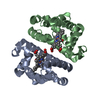 2r4wC 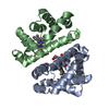 2r4xC 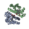 2r4yC 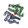 2r4zC 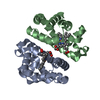 2z85C 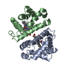 3sdhS S: Starting model for refinement C: citing same article ( |
|---|---|
| Similar structure data |


-Links
 PDBj
PDBj


- Assembly
| Deposited unit | 
| ||||||||
|---|---|---|---|---|---|---|---|---|---|
| 1 |
| ||||||||
| Unit cell |
| ||||||||
| Components on special symmetry positions |
|
-Components
-Protein , 1 types, 2 molecules AB
| #1: Protein | Mass: 16040.356 Da / Num. of mol.: 2 / Mutation: I1025W/I2025W Source method: isolated from a genetically manipulated source Source: (gene. exp.) Scapharca inaequivalvis (ark clam) / Gene: HBI / Plasmid: PCS-26 / Production host:  |
|---|
-Non-polymers , 5 types, 382 molecules 


| #2: Chemical |  Mass: 616.487 Da / Num. of mol.: 2 / Source method: obtained synthetically / Formula: C34H32FeN4O4 Mass: 616.487 Da / Num. of mol.: 2 / Source method: obtained synthetically / Formula: C34H32FeN4O4#3: Chemical |  Mass: 28.010 Da / Num. of mol.: 2 / Source method: obtained synthetically / Formula: CO Mass: 28.010 Da / Num. of mol.: 2 / Source method: obtained synthetically / Formula: CO#4: Chemical | ChemComp-XE /  Mass: 131.293 Da / Num. of mol.: 4 / Source method: obtained synthetically / Formula: Xe Mass: 131.293 Da / Num. of mol.: 4 / Source method: obtained synthetically / Formula: Xe#5: Chemical | ChemComp-PO4 / |  Mass: 94.971 Da / Num. of mol.: 1 / Source method: obtained synthetically / Formula: PO4 Mass: 94.971 Da / Num. of mol.: 1 / Source method: obtained synthetically / Formula: PO4#6: Water | ChemComp-HOH / | Mass: 18.015 Da / Num. of mol.: 373 / Source method: isolated from a natural source / Formula: H2O |
|---|
-Experimental details
-Experiment
| Experiment | Method: X-RAY DIFFRACTION / Number of used crystals: 1 |
|---|
- Sample preparation
| Crystal | Density Matthews: 2.11 Å3/Da / Density % sol: 42 % |
|---|---|
| Crystal grow | Temperature: 298 K / Method: small tubes / pH: 7.5 Details: 1.5-2.5M PHOSPHATE BUFFER, pH 7.50, SMALL TUBES, temperature 298K |
-Data collection
| Diffraction | Mean temperature: 110 K |
|---|---|
| Diffraction source | Source: SYNCHROTRON / Site: APS  / Beamline: 14-BM-C / Wavelength: 0.9 Å / Beamline: 14-BM-C / Wavelength: 0.9 Å |
| Detector | Type: ADSC QUANTUM 315 / Detector: CCD / Date: Nov 7, 2005 |
| Radiation | Protocol: SINGLE WAVELENGTH / Monochromatic (M) / Laue (L): M / Scattering type: x-ray |
| Radiation wavelength | Wavelength: 0.9 Å / Relative weight: 1 |
| Reflection | Resolution: 1.06→46.3 Å / Num. obs: 118984 / % possible obs: 95.3 % / Observed criterion σ(I): 0 / Redundancy: 4.4 % / Rmerge(I) obs: 0.072 / Rsym value: 0.072 / Net I/σ(I): 24.1 |
| Reflection shell | Resolution: 1.06→1.1 Å / Rmerge(I) obs: 0.286 / Mean I/σ(I) obs: 5.5 / Rsym value: 0.286 / % possible all: 90.4 |
- Processing
| Software |
| ||||||||||||||||||||||||
|---|---|---|---|---|---|---|---|---|---|---|---|---|---|---|---|---|---|---|---|---|---|---|---|---|---|
| Refinement | Method to determine structure: MOLECULAR REPLACEMENT Starting model: PDB ENTRY 3SDH Resolution: 1.06→46.3 Å / Isotropic thermal model: ANISOTROPIC / Cross valid method: THROUGHOUT / σ(F): 0 Details: THE MODEL WAS REFINED WITH A COMBINATION OF CNS AND SHELX WITH A ROUND OF MANUAL INTERVENTION IN BETWEEN.
| ||||||||||||||||||||||||
| Refinement step | Cycle: LAST / Resolution: 1.06→46.3 Å
| ||||||||||||||||||||||||
| Refine LS restraints |
| ||||||||||||||||||||||||
| LS refinement shell | Resolution: 1.06→1.1 Å / Total num. of bins used: 10
|
