 Movie
Movie Controller


+ Open data

- Basic information
| Entry | Database: PDB / ID: 2ylb | ||||||
|---|---|---|---|---|---|---|---|
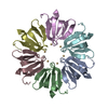
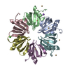
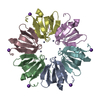
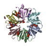
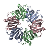
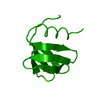
| Title | Structure of Salmonella typhimurium Hfq at 1.15 A | ||||||
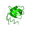
 Components Components | PROTEIN HFQ | ||||||
 Keywords Keywords | RNA BINDING PROTEIN / RNA-BINDING PROTEIN / LSM PROTEIN / RNA CHAPERONE | ||||||
| Function / homology |  Function and homology information Function and homology informationregulation of translation, ncRNA-mediated / regulation of RNA stability / regulation of DNA-templated transcription / RNA binding / identical protein binding / cytosol Similarity search - Function | ||||||
| Biological species |  SALMONELLA ENTERICA SUBSP. ENTERICA SEROVAR TYPHIMURIUM (bacteria) SALMONELLA ENTERICA SUBSP. ENTERICA SEROVAR TYPHIMURIUM (bacteria) | ||||||
| Method |  X-RAY DIFFRACTION / SYNCHROTRON / MOLECULAR REPLACEMENT / Resolution: 1.15 Å X-RAY DIFFRACTION / SYNCHROTRON / MOLECULAR REPLACEMENT / Resolution: 1.15 Å | ||||||
 Authors Authors | Sauer, E. / Weichenrieder, O. | ||||||
 Citation Citation | Journal: Proc.Natl.Acad.Sci.USA / Year: 2011 Title: Structural Basis for RNA 3' End Recognition by Hfq Authors: Sauer, E. / Weichenrieder, O. | ||||||
| History |
| ||||||
| Remark 650 | HELIX DETERMINATION METHOD: AUTHOR PROVIDED. | ||||||
| Remark 700 | SHEET DETERMINATION METHOD: DSSP THE SHEETS PRESENTED AS "AA" IN EACH CHAIN ON SHEET RECORDS BELOW ... SHEET DETERMINATION METHOD: DSSP THE SHEETS PRESENTED AS "AA" IN EACH CHAIN ON SHEET RECORDS BELOW IS ACTUALLY AN 30-STRANDED BARREL THIS IS REPRESENTED BY A 31-STRANDED SHEET IN WHICH THE FIRST AND LAST STRANDS ARE IDENTICAL. |
- Structure visualization
| Structure viewer | Molecule: MolmilJmol/JSmol |
|---|
- Downloads & links
-Download
| PDBx/mmCIF format | 2ylb.cif.gz | 262.5 KB | Display | PDBx/mmCIF format |
|---|---|---|---|---|
| PDB format | pdb2ylb.ent.gz | 218.5 KB | Display | PDB format |
| PDBx/mmJSON format | 2ylb.json.gz | Tree view | PDBx/mmJSON format | |
| Others |  Other downloads Other downloads |
-Validation report
| Arichive directory | https://data.pdbj.org/pub/pdb/validation_reports/yl/2ylbftp://data.pdbj.org/pub/pdb/validation_reports/yl/2ylb | HTTPS FTP |
|---|
-Related structure data
| Related structure data | 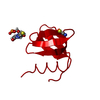 2ylcC 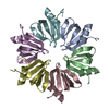 1hk9S C: citing same article ( S: Starting model for refinement |
|---|---|
| Similar structure data |


-Links
 PDBj
PDBj

- Assembly
| Deposited unit | 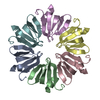
| ||||||||
|---|---|---|---|---|---|---|---|---|---|
| 1 |
| ||||||||
| Unit cell |
|
-Components
| #1: Protein | Mass: 8257.572 Da / Num. of mol.: 6 / Fragment: RESIDUES 1-72 Source method: isolated from a genetically manipulated source Details: THE SEQUENCE IS PRECEDED BY A GA TAG REMAINING FROM THE PURIFICATION TAG. Source: (gene. exp.) SALMONELLA ENTERICA SUBSP. ENTERICA SEROVAR TYPHIMURIUM (bacteria)Strain: LT2 / Plasmid: PET M60 / Production host: #2: Water | ChemComp-HOH / |  Mass: 18.015 Da / Num. of mol.: 376 / Source method: isolated from a natural source / Formula: H2O Mass: 18.015 Da / Num. of mol.: 376 / Source method: isolated from a natural source / Formula: H2OSequence details | THE SEQUENCE IS PRECEDED BY A GA TAG REMAINING FROM THE PURIFICATI | |
|---|
-Experimental details
-Experiment
| Experiment | Method: X-RAY DIFFRACTION / Number of used crystals: 1 |
|---|
- Sample preparation
| Crystal | Density Matthews: 2 Å3/Da / Density % sol: 39 % / Description: NONE |
|---|---|
| Crystal grow | pH: 7 Details: 0.1 M HEPES (PH=7.0), 0.5 % JEFFAMINE, 1.1 M MALONATE |
-Data collection
| Diffraction | Mean temperature: 90 K |
|---|---|
| Diffraction source | Source: SYNCHROTRON / Site: SLS  / Beamline: X10SA / Wavelength: 0.827 / Beamline: X10SA / Wavelength: 0.827 |
| Detector | Type: MARRESEARCH / Detector: CCD / Date: May 19, 2009 / Details: MIRRORS |
| Radiation | Monochromator: SI(111) / Protocol: SINGLE WAVELENGTH / Monochromatic (M) / Laue (L): M / Scattering type: x-ray |
| Radiation wavelength | Wavelength: 0.827 Å / Relative weight: 1 |
| Reflection | Resolution: 1.15→20 Å / Num. obs: 123284 / % possible obs: 98 % / Observed criterion σ(I): -3 / Redundancy: 4.9 % / Biso Wilson estimate: 8.2 Å2 / Rsym value: 0.067 / Net I/σ(I): 12.8 |
| Reflection shell | Resolution: 1.15→1.18 Å / Redundancy: 3.9 % / Mean I/σ(I) obs: 2.52 / Rsym value: 0.64 / % possible all: 96.9 |
- Processing
| Software |
| |||||||||||||||||||||||||||||||||||||||||||||||||||||||||||||||||||||||||||||
|---|---|---|---|---|---|---|---|---|---|---|---|---|---|---|---|---|---|---|---|---|---|---|---|---|---|---|---|---|---|---|---|---|---|---|---|---|---|---|---|---|---|---|---|---|---|---|---|---|---|---|---|---|---|---|---|---|---|---|---|---|---|---|---|---|---|---|---|---|---|---|---|---|---|---|---|---|---|---|
| Refinement | Method to determine structure: MOLECULAR REPLACEMENT Starting model: PDB ENTRY 1HK9 Resolution: 1.15→18.84 Å / SU ML: 0.12 / σ(F): 1.99 / Phase error: 21.88 / Stereochemistry target values: ML Details: HYDROGENS WERE REFINED IN THE RIDING POSITIONS. B FACTORS WERE REFINED ANISOTROPICALLY FOR NON-HYDROGEN ATOMS. THE FOLLOWING RESIDUES WERE MODELED AS DOUBLE CONFORMATIONS. CHAIN A, RESIDUES ...Details: HYDROGENS WERE REFINED IN THE RIDING POSITIONS. B FACTORS WERE REFINED ANISOTROPICALLY FOR NON-HYDROGEN ATOMS. THE FOLLOWING RESIDUES WERE MODELED AS DOUBLE CONFORMATIONS. CHAIN A, RESIDUES 60, 66. CHAIN B, RESIDUES 17, 19, 36, 38, 60, 66. CHAIN C, RESIDUES 17, 38, 60, 61, 66. CHAIN D, RESIDUES 13, 36. CHAIN E, RESIDUES 36, 66. CHAIN F, RESIDUE 61.THE FOLLOWING RESIDUES ARE DISORDERED. CHAIN A, RESIDUES 1 TO 5, 71 TO 72. CHAIN B, RESIDUES 1 TO 6, 72. CHAIN C, RESIDUES 1 TO 3, 72. CHAIN D, RESIDUES 1 TO 3, 72. CHAIN E, RESIDUES 1 TO 3, 72. CHAIN F, RESIDUES 1 TO 5, 72.
| |||||||||||||||||||||||||||||||||||||||||||||||||||||||||||||||||||||||||||||
| Solvent computation | Shrinkage radii: 0.89 Å / VDW probe radii: 1 Å / Solvent model: FLAT BULK SOLVENT MODEL / Bsol: 45.557 Å2 / ksol: 0.461 e/Å3 | |||||||||||||||||||||||||||||||||||||||||||||||||||||||||||||||||||||||||||||
| Displacement parameters | Biso mean: 15.6 Å2
| |||||||||||||||||||||||||||||||||||||||||||||||||||||||||||||||||||||||||||||
| Refinement step | Cycle: LAST / Resolution: 1.15→18.84 Å
| |||||||||||||||||||||||||||||||||||||||||||||||||||||||||||||||||||||||||||||
| Refine LS restraints |
| |||||||||||||||||||||||||||||||||||||||||||||||||||||||||||||||||||||||||||||
| LS refinement shell |
|
