 Movie
Movie Controller

[English] 日本語
 Yorodumi
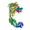
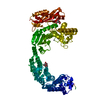
- PDB-2y3u: Crystal structure of apo collagenase G from Clostridium histolyti... -
+ Open data

- Basic information
| Entry | Database: PDB / ID: 2y3u | ||||||
|---|---|---|---|---|---|---|---|
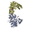
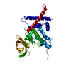
| Title | Crystal structure of apo collagenase G from Clostridium histolyticum at 2.55 Angstrom resolution | ||||||
 Components Components | COLLAGENASE | ||||||
 Keywords Keywords | HYDROLASE / GLUZINCIN / METALLOPROTEASE | ||||||
| Function / homology |  Function and homology informationtripeptidase activity / microbial collagenase / collagen metabolic process / collagen binding / metalloendopeptidase activity / endopeptidase activity / calcium ion binding / proteolysis / zinc ion binding / extracellular region Function and homology informationtripeptidase activity / microbial collagenase / collagen metabolic process / collagen binding / metalloendopeptidase activity / endopeptidase activity / calcium ion binding / proteolysis / zinc ion binding / extracellular regionSimilarity search - Function | ||||||
| Biological species |  CLOSTRIDIUM HISTOLYTICUM (bacteria) CLOSTRIDIUM HISTOLYTICUM (bacteria) | ||||||
| Method | X-RAY DIFFRACTION / SYNCHROTRON / MAD / Resolution: 2.55 Å | ||||||
 Authors Authors | Eckhard, U. / Brandstetter, H. | ||||||
 Citation Citation | Journal: Nat.Struct.Mol.Biol. / Year: 2011 Title: Structure of Collagenase G Reveals a Chew-and -Digest Mechanism of Bacterial Collagenolysis Authors: Eckhard, U. / Schoenauer, E. / Nuess, D. / Brandstetter, H. | ||||||
| History |
|
- Structure visualization
| Structure viewer | Molecule: MolmilJmol/JSmol |
|---|
- Downloads & links
-Download
| PDBx/mmCIF format | 2y3u.cif.gz | 289.2 KB | Display | PDBx/mmCIF format |
|---|---|---|---|---|
| PDB format | pdb2y3u.ent.gz | 244.7 KB | Display | PDB format |
| PDBx/mmJSON format | 2y3u.json.gz | Tree view | PDBx/mmJSON format | |
| Others |  Other downloads Other downloads |
-Validation report
| Arichive directory | https://data.pdbj.org/pub/pdb/validation_reports/y3/2y3uftp://data.pdbj.org/pub/pdb/validation_reports/y3/2y3u | HTTPS FTP |
|---|
-Related structure data


-Links
 PDBj
PDBj


- Assembly
| Deposited unit | 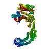
| ||||||||
|---|---|---|---|---|---|---|---|---|---|
| 1 |
| ||||||||
| Unit cell |
|
-Components
| #1: Protein | / COLLAGENASE G Mass: 89594.695 Da / Num. of mol.: 1 / Fragment: RESIDUES 119-880 Source method: isolated from a genetically manipulated source Source: (gene. exp.) CLOSTRIDIUM HISTOLYTICUM (bacteria) / Production host: ESCHERICHIA COLI (E. coli) / Strain (production host): B834 / References: UniProt: Q9X721, microbial collagenase |
|---|---|
| #2: Chemical | ChemComp-P6G / Polyethylene glycol  Mass: 282.331 Da / Num. of mol.: 1 / Source method: obtained synthetically / Formula: C12H26O7 / Comment: precipitant*YM Mass: 282.331 Da / Num. of mol.: 1 / Source method: obtained synthetically / Formula: C12H26O7 / Comment: precipitant*YM |
| #3: Chemical | ChemComp-FLC / Citric acid  Mass: 189.100 Da / Num. of mol.: 1 / Source method: obtained synthetically / Formula: C6H5O7 Mass: 189.100 Da / Num. of mol.: 1 / Source method: obtained synthetically / Formula: C6H5O7 |
| #4: Chemical | ChemComp-PEG / Diethylene glycol  Mass: 106.120 Da / Num. of mol.: 1 / Source method: obtained synthetically / Formula: C4H10O3 Mass: 106.120 Da / Num. of mol.: 1 / Source method: obtained synthetically / Formula: C4H10O3 |
| #5: Water | ChemComp-HOH / Water Mass: 18.015 Da / Num. of mol.: 86 / Source method: isolated from a natural source / Formula: H2O Mass: 18.015 Da / Num. of mol.: 86 / Source method: isolated from a natural source / Formula: H2O |
| Nonpolymer details | CATALYTIC ZN BINDING SITE IS FORMED BY HIS523, HIS527 AND GLU555. ZN IS REPLACED BY WATER A2086 IN ...CATALYTIC ZN BINDING SITE IS FORMED BY HIS523, HIS527 AND GLU555. ZN IS REPLACED BY WATER A2086 IN THIS STRUCTURE |
| Sequence details | NATURAL VARIATION AT THESE THREE POSITIONS OWING TO ISOLATION FROM A DIFFERENT STRAIN. |
-Experimental details
-Experiment
| Experiment | Method: X-RAY DIFFRACTION / Number of used crystals: 1 |
|---|
- Sample preparation
| Crystal | Density Matthews: 3.15 Å3/Da / Density % sol: 61 % / Description: NONE |
|---|---|
| Crystal grow | pH: 8.3 / Details: 21.5% PEG 3350, 0.225 M TRISODIUM CITRATE PH 8.3. |
-Data collection
| Diffraction | Mean temperature: 100 K |
|---|---|
| Diffraction source | Source: SYNCHROTRON / Site: BESSY  / Beamline: 14.1 / Wavelength: 0.98793 / Beamline: 14.1 / Wavelength: 0.98793 |
| Detector | Type: MARMOSAIC 225 mm CCD / Detector: CCD / Date: Aug 12, 2010 / Details: MIRRORS |
| Radiation | Monochromator: DOUBLE CRYSTAL / Protocol: MAD / Monochromatic (M) / Laue (L): M / Scattering type: x-ray |
| Radiation wavelength | Wavelength: 0.98793 Å / Relative weight: 1 |
| Reflection | Resolution: 2.55→48.32 Å / Num. obs: 33182 / % possible obs: 88.1 % / Observed criterion σ(I): 1.8 / Redundancy: 5.4 % / Biso Wilson estimate: 62.2 Å2 / Rmerge(I) obs: 0.1 / Net I/σ(I): 9.3 |
| Reflection shell | Resolution: 2.55→2.69 Å / Redundancy: 5 % / Rmerge(I) obs: 0.7 / Mean I/σ(I) obs: 1.8 / % possible all: 84.4 |
- Processing
| Software |
| ||||||||||||||||||||||||||||||||||||||||||||||||||||||||||||||||||||||||||||||||||||||||||||||||||||||||||||||||||||||||||||||||||||||||||||||||||||||||||||||||||||||||||||||||||||||
|---|---|---|---|---|---|---|---|---|---|---|---|---|---|---|---|---|---|---|---|---|---|---|---|---|---|---|---|---|---|---|---|---|---|---|---|---|---|---|---|---|---|---|---|---|---|---|---|---|---|---|---|---|---|---|---|---|---|---|---|---|---|---|---|---|---|---|---|---|---|---|---|---|---|---|---|---|---|---|---|---|---|---|---|---|---|---|---|---|---|---|---|---|---|---|---|---|---|---|---|---|---|---|---|---|---|---|---|---|---|---|---|---|---|---|---|---|---|---|---|---|---|---|---|---|---|---|---|---|---|---|---|---|---|---|---|---|---|---|---|---|---|---|---|---|---|---|---|---|---|---|---|---|---|---|---|---|---|---|---|---|---|---|---|---|---|---|---|---|---|---|---|---|---|---|---|---|---|---|---|---|---|---|---|
| Refinement | Method to determine structure: MAD Starting model: NONE Resolution: 2.55→40 Å / Cor.coef. Fo:Fc: 0.949 / Cor.coef. Fo:Fc free: 0.919 / SU B: 25.2 / SU ML: 0.238 / Cross valid method: THROUGHOUT / ESU R: 0.429 / ESU R Free: 0.291 / Stereochemistry target values: MAXIMUM LIKELIHOOD / Details: HYDROGENS HAVE BEEN ADDED IN THE RIDING POSITIONS.
| ||||||||||||||||||||||||||||||||||||||||||||||||||||||||||||||||||||||||||||||||||||||||||||||||||||||||||||||||||||||||||||||||||||||||||||||||||||||||||||||||||||||||||||||||||||||
| Solvent computation | Ion probe radii: 0.8 Å / Shrinkage radii: 0.8 Å / VDW probe radii: 1.4 Å / Solvent model: MASK | ||||||||||||||||||||||||||||||||||||||||||||||||||||||||||||||||||||||||||||||||||||||||||||||||||||||||||||||||||||||||||||||||||||||||||||||||||||||||||||||||||||||||||||||||||||||
| Displacement parameters | Biso mean: 58.695 Å2
| ||||||||||||||||||||||||||||||||||||||||||||||||||||||||||||||||||||||||||||||||||||||||||||||||||||||||||||||||||||||||||||||||||||||||||||||||||||||||||||||||||||||||||||||||||||||
| Refinement step | Cycle: LAST / Resolution: 2.55→40 Å
| ||||||||||||||||||||||||||||||||||||||||||||||||||||||||||||||||||||||||||||||||||||||||||||||||||||||||||||||||||||||||||||||||||||||||||||||||||||||||||||||||||||||||||||||||||||||
| Refine LS restraints |
|