 Movie
Movie Controller

[English] 日本語
 Yorodumi
- PDB-2y72: Crystal structure of the PKD Domain of Collagenase G from Clostri... -
+ Open data

- Basic information
| Entry | Database: PDB / ID: 2y72 | ||||||
|---|---|---|---|---|---|---|---|
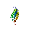
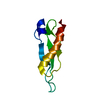
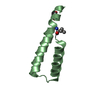
| Title | Crystal structure of the PKD Domain of Collagenase G from Clostridium Histolyticum at 1.18 Angstrom Resolution. | ||||||
 Components Components | COLLAGENASE | ||||||
 Keywords Keywords | HYDROLASE / POLYCYSTIC KIDNEY DISEASE DOMAIN / BETA BARREL / COLLAGEN RECOGNITION DOMAIN | ||||||
| Function / homology |  Function and homology information Function and homology informationtripeptidase activity / microbial collagenase / collagen metabolic process / collagen binding / metalloendopeptidase activity / endopeptidase activity / calcium ion binding / proteolysis / extracellular region / zinc ion binding Similarity search - Function | ||||||
| Biological species |  CLOSTRIDIUM HISTOLYTICUM (bacteria) CLOSTRIDIUM HISTOLYTICUM (bacteria) | ||||||
| Method |  X-RAY DIFFRACTION / SYNCHROTRON / MOLECULAR REPLACEMENT / Resolution: 1.18 Å X-RAY DIFFRACTION / SYNCHROTRON / MOLECULAR REPLACEMENT / Resolution: 1.18 Å | ||||||
 Authors Authors | Eckhard, U. / Brandstetter, H. | ||||||
 Citation Citation | Journal: Nat.Struct.Mol.Biol. / Year: 2011 Title: Structure of Collagenase G Reveals a Chew-and -Digest Mechanism of Bacterial Collagenolysis Authors: Eckhard, U. / Schoenauer, E. / Nuess, D. / Brandstetter, H. #1: Journal: Biol.Chem / Year: 2011 Title: Polycystic Kidney Disease-Like Domains of Clostridial Collagenases and Their Role in Collagen Recruitment. Authors: Eckhard, U. / Brandstetter, H. | ||||||
| History |
|
- Structure visualization
| Structure viewer | Molecule: MolmilJmol/JSmol |
|---|
- Downloads & links
-Download
| PDBx/mmCIF format | 2y72.cif.gz | 94.5 KB | Display | PDBx/mmCIF format |
|---|---|---|---|---|
| PDB format | pdb2y72.ent.gz | 73.7 KB | Display | PDB format |
| PDBx/mmJSON format | 2y72.json.gz | Tree view | PDBx/mmJSON format | |
| Others |  Other downloads Other downloads |
-Validation report
| Arichive directory | https://data.pdbj.org/pub/pdb/validation_reports/y7/2y72ftp://data.pdbj.org/pub/pdb/validation_reports/y7/2y72 | HTTPS FTP |
|---|
-Related structure data
| Related structure data |  2y3uC 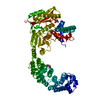 2y50C  2y6iC  2c26S C: citing same article ( S: Starting model for refinement |
|---|---|
| Similar structure data |


-Links
 PDBj
PDBj- Assembly
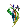
| Deposited unit | 
| ||||||||
|---|---|---|---|---|---|---|---|---|---|
| 1 | 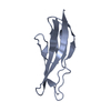
| ||||||||
| 2 | 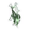
| ||||||||
| Unit cell |
|
-Components
| #1: Protein | Mass: 8946.764 Da / Num. of mol.: 2 Fragment: POLYCYSTIC KIDNEY DISEASE (PKD) DOMAIN, RESIDUES 799-880 Source method: isolated from a genetically manipulated source Source: (gene. exp.) CLOSTRIDIUM HISTOLYTICUM (bacteria) / Production host: #2: Water | ChemComp-HOH / |  Mass: 18.015 Da / Num. of mol.: 364 / Source method: isolated from a natural source / Formula: H2O Mass: 18.015 Da / Num. of mol.: 364 / Source method: isolated from a natural source / Formula: H2O |
|---|
-Experimental details
-Experiment
| Experiment | Method: X-RAY DIFFRACTION / Number of used crystals: 2 |
|---|
- Sample preparation
| Crystal | Density Matthews: 2.55 Å3/Da / Density % sol: 51.74 % / Description: NONE |
|---|---|
| Crystal grow | pH: 7 / Details: 2.5M C3H2O4NA2, 10MM CACL2, PH 7.5 |
-Data collection
| Diffraction | Mean temperature: 100 K |
|---|---|
| Diffraction source | Source: SYNCHROTRON / Site: BESSY  / Beamline: 14.1 / Wavelength: 0.98793 / Beamline: 14.1 / Wavelength: 0.98793 |
| Detector | Type: MARRESEARCH / Detector: CCD / Date: Aug 12, 2010 / Details: MIRRORS |
| Radiation | Monochromator: DOUBLE CRYSTAL / Protocol: SINGLE WAVELENGTH / Monochromatic (M) / Laue (L): M / Scattering type: x-ray |
| Radiation wavelength | Wavelength: 0.98793 Å / Relative weight: 1 |
| Reflection | Resolution: 1.18→40.51 Å / Num. obs: 56646 / % possible obs: 96.4 % / Observed criterion σ(I): 3.2 / Redundancy: 6 % / Biso Wilson estimate: 9.7 Å2 / Rmerge(I) obs: 0.07 / Net I/σ(I): 13.9 |
| Reflection shell | Resolution: 1.18→1.24 Å / Redundancy: 2.9 % / Rmerge(I) obs: 0.3 / Mean I/σ(I) obs: 3.2 / % possible all: 78.4 |
- Processing
| Software |
| ||||||||||||||||||||||||||||||||||||||||||||||||||||||||||||||||||||||||||||||||||||||||||||||||||||||||||||||||||||||||||||||||||||||||||||||||||||||||||||||||||||||||||||||||||||||
|---|---|---|---|---|---|---|---|---|---|---|---|---|---|---|---|---|---|---|---|---|---|---|---|---|---|---|---|---|---|---|---|---|---|---|---|---|---|---|---|---|---|---|---|---|---|---|---|---|---|---|---|---|---|---|---|---|---|---|---|---|---|---|---|---|---|---|---|---|---|---|---|---|---|---|---|---|---|---|---|---|---|---|---|---|---|---|---|---|---|---|---|---|---|---|---|---|---|---|---|---|---|---|---|---|---|---|---|---|---|---|---|---|---|---|---|---|---|---|---|---|---|---|---|---|---|---|---|---|---|---|---|---|---|---|---|---|---|---|---|---|---|---|---|---|---|---|---|---|---|---|---|---|---|---|---|---|---|---|---|---|---|---|---|---|---|---|---|---|---|---|---|---|---|---|---|---|---|---|---|---|---|---|---|
| Refinement | Method to determine structure: MOLECULAR REPLACEMENT Starting model: PDB ENTRY 2C26 Resolution: 1.18→44.94 Å / Cor.coef. Fo:Fc: 0.975 / Cor.coef. Fo:Fc free: 0.964 / SU B: 1.108 / SU ML: 0.023 / Cross valid method: THROUGHOUT / ESU R: 0.037 / ESU R Free: 0.039 / Stereochemistry target values: MAXIMUM LIKELIHOOD / Details: HYDROGENS HAVE BEEN ADDED IN THE RIDING POSITIONS.
| ||||||||||||||||||||||||||||||||||||||||||||||||||||||||||||||||||||||||||||||||||||||||||||||||||||||||||||||||||||||||||||||||||||||||||||||||||||||||||||||||||||||||||||||||||||||
| Solvent computation | Ion probe radii: 0.8 Å / Shrinkage radii: 0.8 Å / VDW probe radii: 1.4 Å / Solvent model: MASK | ||||||||||||||||||||||||||||||||||||||||||||||||||||||||||||||||||||||||||||||||||||||||||||||||||||||||||||||||||||||||||||||||||||||||||||||||||||||||||||||||||||||||||||||||||||||
| Displacement parameters | Biso mean: 12.061 Å2
| ||||||||||||||||||||||||||||||||||||||||||||||||||||||||||||||||||||||||||||||||||||||||||||||||||||||||||||||||||||||||||||||||||||||||||||||||||||||||||||||||||||||||||||||||||||||
| Refinement step | Cycle: LAST / Resolution: 1.18→44.94 Å
| ||||||||||||||||||||||||||||||||||||||||||||||||||||||||||||||||||||||||||||||||||||||||||||||||||||||||||||||||||||||||||||||||||||||||||||||||||||||||||||||||||||||||||||||||||||||
| Refine LS restraints |
|