 Movie
Movie Controller

[English] 日本語
 Yorodumi
- PDB-3bhp: Crystal structure of UPF0291 protein ynzC from Bacillus subtilis ... -
+ Open data

- Basic information
| Entry | Database: PDB / ID: 3bhp | ||||||
|---|---|---|---|---|---|---|---|
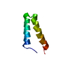
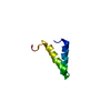
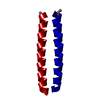
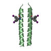
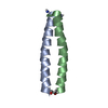
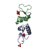
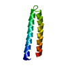
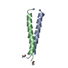
| Title | Crystal structure of UPF0291 protein ynzC from Bacillus subtilis at resolution 2.0 A. Northeast Structural Genomics Consortium target SR384 | ||||||
 Components Components | UPF0291 protein ynzC | ||||||
 Keywords Keywords | STRUCTURAL GENOMICS / UNKNOWN FUNCTION / NESG / SR384 / O31818 / UPF0291 protein ynzC / PSI-2 / Protein Structure Initiative / Northeast Structural Genomics Consortium / Cytoplasm | ||||||
| Function / homology | Protein of unknown function DUF896 / Bacterial protein of unknown function (DUF896) / Helix hairpin bin / Helix Hairpins / Orthogonal Bundle / Mainly Alpha / cytoplasm / UPF0291 protein YnzC Function and homology information Function and homology information | ||||||
| Biological species |  | ||||||
| Method |  X-RAY DIFFRACTION / SYNCHROTRON / SAD / Resolution: 2.01 Å X-RAY DIFFRACTION / SYNCHROTRON / SAD / Resolution: 2.01 Å | ||||||
 Authors Authors | Kuzin, A.P. / Su, M. / Seetharaman, J. / Janjua, H. / Cunningham, K. / Maglaqui, M. / Owens, L.A. / Zhao, L. / Xiao, R. / Baran, M.C. ...Kuzin, A.P. / Su, M. / Seetharaman, J. / Janjua, H. / Cunningham, K. / Maglaqui, M. / Owens, L.A. / Zhao, L. / Xiao, R. / Baran, M.C. / Acton, T.B. / Rost, B. / Montelione, G.T. / Hunt, J.F. / Tong, L. / Northeast Structural Genomics Consortium (NESG) | ||||||
 Citation Citation | Journal: To be Published Title: Crystal structure of the UPF0291 protein ynzC from Bacillus subtilis at the resolution 2.0 A. Authors: Kuzin, A.P. / Su, M. / Seetharaman, J. / Janjua, H. / Cunningham, K. / Maglaqui, M. / Owens, L.A. / Zhao, L. / Xiao, R. / Baran, M.C. / Acton, T.B. / Rost, B. / Montelione, G.T. / Hunt, J.F. / Tong, L. | ||||||
| History |
|
- Structure visualization
| Structure viewer | Molecule: MolmilJmol/JSmol |
|---|
- Downloads & links
-Download
| PDBx/mmCIF format | 3bhp.cif.gz | 45.5 KB | Display | PDBx/mmCIF format |
|---|---|---|---|---|
| PDB format | pdb3bhp.ent.gz | 33.3 KB | Display | PDB format |
| PDBx/mmJSON format | 3bhp.json.gz | Tree view | PDBx/mmJSON format | |
| Others |  Other downloads Other downloads |
-Validation report
| Arichive directory | https://data.pdbj.org/pub/pdb/validation_reports/bh/3bhpftp://data.pdbj.org/pub/pdb/validation_reports/bh/3bhp | HTTPS FTP |
|---|
-Related structure data
| Related structure data | |
|---|---|
| Similar structure data | |
| Other databases |


-Links
 PDBj
PDBj- Assembly
| Deposited unit | 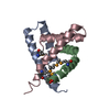
| ||||||||
|---|---|---|---|---|---|---|---|---|---|
| 1 | 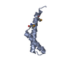
| ||||||||
| 2 | 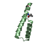
| ||||||||
| 3 | 
| ||||||||
| 4 |
| ||||||||
| Unit cell |
| ||||||||
| Components on special symmetry positions |
|
-Components
| #1: Protein | Mass: 7027.784 Da / Num. of mol.: 3 / Fragment: Residues 1-52 Source method: isolated from a genetically manipulated source Source: (gene. exp.) #2: Water | ChemComp-HOH / |  Mass: 18.015 Da / Num. of mol.: 196 / Source method: isolated from a natural source / Formula: H2O Mass: 18.015 Da / Num. of mol.: 196 / Source method: isolated from a natural source / Formula: H2OHas protein modification | Y | |
|---|
-Experimental details
-Experiment
| Experiment | Method: X-RAY DIFFRACTION / Number of used crystals: 1 |
|---|
- Sample preparation
| Crystal | Density Matthews: 2.46 Å3/Da / Density % sol: 49.96 % Description: The structure factor file contains Friedel pairs |
|---|---|
| Crystal grow | Temperature: 293 K / Method: vapor diffusion, hanging drop / pH: 4 Details: 0.1M KH2PO4, 0.1M Na Citrate, 40% PEG 1000, pH 4.0, VAPOR DIFFUSION, HANGING DROP, temperature 293K |
-Data collection
| Diffraction | Mean temperature: 100 K |
|---|---|
| Diffraction source | Source: SYNCHROTRON / Site: NSLS  / Beamline: X4A / Wavelength: 0.979 Å / Beamline: X4A / Wavelength: 0.979 Å |
| Detector | Type: ADSC QUANTUM 210 / Detector: CCD / Date: Oct 9, 2007 |
| Radiation | Protocol: SINGLE WAVELENGTH / Monochromatic (M) / Laue (L): M / Scattering type: x-ray |
| Radiation wavelength | Wavelength: 0.979 Å / Relative weight: 1 |
| Reflection | Resolution: 2→30 Å / Num. obs: 26190 / % possible obs: 98.9 % / Observed criterion σ(I): -3 / Redundancy: 15.5 % / Biso Wilson estimate: 7.1 Å2 / Rmerge(I) obs: 0.058 / Net I/σ(I): 25.4 |
| Reflection shell | Resolution: 2→2.07 Å / Rmerge(I) obs: 0.123 / Mean I/σ(I) obs: 10.7 / % possible all: 99.2 |
- Processing
| Software |
| ||||||||||||||||||||||||||||||||||||
|---|---|---|---|---|---|---|---|---|---|---|---|---|---|---|---|---|---|---|---|---|---|---|---|---|---|---|---|---|---|---|---|---|---|---|---|---|---|
| Refinement | Method to determine structure: SAD / Resolution: 2.01→19.55 Å / Rfactor Rfree error: 0.005 / Data cutoff high absF: 660559.39 / Data cutoff low absF: 0 / Isotropic thermal model: RESTRAINED / Cross valid method: THROUGHOUT / σ(F): 1 / Stereochemistry target values: Engh & Huber Details: The Friedel pairs were used in phasing. BULK SOLVENT MODEL USED IN REFINEMENT
| ||||||||||||||||||||||||||||||||||||
| Solvent computation | Solvent model: FLAT MODEL / Bsol: 62.6638 Å2 / ksol: 0.45 e/Å3 | ||||||||||||||||||||||||||||||||||||
| Displacement parameters | Biso mean: 25.9 Å2
| ||||||||||||||||||||||||||||||||||||
| Refine analyze |
| ||||||||||||||||||||||||||||||||||||
| Refinement step | Cycle: LAST / Resolution: 2.01→19.55 Å
| ||||||||||||||||||||||||||||||||||||
| Refine LS restraints |
| ||||||||||||||||||||||||||||||||||||
| LS refinement shell | Resolution: 2.01→2.13 Å / Rfactor Rfree error: 0.012 / Total num. of bins used: 6
| ||||||||||||||||||||||||||||||||||||
| Xplor file |
|
