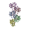
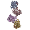
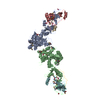
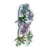
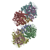
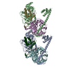
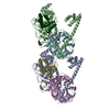
The As.u. contains a monomer but the Biological unit is a dimer. The second part of the biological assembly is generated by applying the symetry operation -x, y, -z and the translation 1 0 1
-
Components
#1: Protein
Phosphoenolpyruvate-proteinphosphotransferase / E.C.2.7.3.9 / Phosphotransferase system / enzyme I / PEP-dependent HPr protein kinase phosphoryltransferase
Mass: 63361.570 Da / Num. of mol.: 1 Source method: isolated from a genetically manipulated source Source: (gene. exp.) Staphylococcus carnosus (bacteria) / Gene: Pts1 / Plasmid: pUC-ptsO2.6X / Production host: Escherichia coli (E. coli) / Strain (production host): DH5a References: UniProt: P23533, phosphoenolpyruvate-protein phosphotransferase
Mass: 18.015 Da / Num. of mol.: 172 / Source method: isolated from a natural source / Formula: H2O
-
Experimental details
-
Experiment
Experiment
Method: X-RAY DIFFRACTION / Number of used crystals: 2
-
Sample preparation
Crystal
ID
Density Matthews (Å3/Da)
Density % sol (%)
1
2.68
54.07
2
Crystal grow
Temperature (K)
Crystal-ID
Method
pH
Details
293
1
vapor diffusion, hanging drop
8.5
30% PEG 4000, 0.2 M Li2SO4 and 0.1 M Tris-HCl pH8.5 and 0.2 l of additive solution (solution No 11 of Hampton Crystal Screen, Hampton Research, VAPOR DIFFUSION, HANGING DROP, temperature 293K
293
2
vapor diffusion, sitting drop
8.5
30% PEG 4000, 0.2 sodium malonate and 0.1 M Tris-HCl at pH of 8.5, VAPOR DIFFUSION, SITTING DROP, temperature 293K
In the structure databanks used in Yorodumi, some data are registered as the other names, "COVID-19 virus" and "2019-nCoV". Here are the details of the virus and the list of structure data.
Jan 31, 2019. EMDB accession codes are about to change! (news from PDBe EMDB page)
EMDB accession codes are about to change! (news from PDBe EMDB page)
The allocation of 4 digits for EMDB accession codes will soon come to an end. Whilst these codes will remain in use, new EMDB accession codes will include an additional digit and will expand incrementally as the available range of codes is exhausted. The current 4-digit format prefixed with “EMD-” (i.e. EMD-XXXX) will advance to a 5-digit format (i.e. EMD-XXXXX), and so on. It is currently estimated that the 4-digit codes will be depleted around Spring 2019, at which point the 5-digit format will come into force.
The EM Navigator/Yorodumi systems omit the EMD- prefix.
Related info.:Q: What is EMD? / ID/Accession-code notation in Yorodumi/EM Navigator
Yorodumi is a browser for structure data from EMDB, PDB, SASBDB, etc.
This page is also the successor to EM Navigator detail page, and also detail information page/front-end page for Omokage search.
The word "yorodu" (or yorozu) is an old Japanese word meaning "ten thousand". "mi" (miru) is to see.
Related info.:EMDB / PDB / SASBDB / Comparison of 3 databanks / Yorodumi Search / Aug 31, 2016. New EM Navigator & Yorodumi / Yorodumi Papers / Jmol/JSmol / Function and homology information / Changes in new EM Navigator and Yorodumi
 Movie
Movie Controller

 Yorodumi
 Open data
 Basic information
 Components
 Keywords
 Function and homology information
 Staphylococcus carnosus (bacteria)
 X-RAY DIFFRACTION /
  Authors
 Citation
 Structure visualization
 Downloads & links
 Other downloads
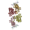
 PDBj
PDBj
 Assembly


 Mass: 96.063 Da / Num. of mol.: 1 / Source method: obtained synthetically / Formula: SO4
 Mass: 18.015 Da / Num. of mol.: 172 / Source method: isolated from a natural source / Formula: H2O
 Sample preparation
 Processing