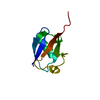
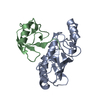
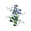
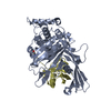
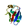
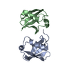
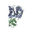
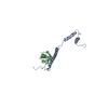
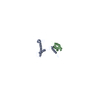
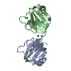
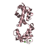
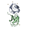
- PDB-2xk5: Crystal structure of K6-linked diubiquitin -
+
Open data
ID or keywords:
Loading...
-
Basic information
Entry
Database: PDB / ID: 2xk5
Title
Crystal structure of K6-linked diubiquitin
Components
(UBIQUITIN) x 2
Keywords
PROTEIN BINDING / SIGNAL TRANSDUCTION / BRCA1
Function / homology
Function and homology information
: / : / protein modification process => GO:0036211 / Formation of the ternary complex, and subsequently, the 43S complex / Ribosomal scanning and start codon recognition / Translation initiation complex formation / SARS-CoV-1 modulates host translation machinery / Peptide chain elongation / Selenocysteine synthesis / Formation of a pool of free 40S subunits ...: / : / protein modification process => GO:0036211 / Formation of the ternary complex, and subsequently, the 43S complex / Ribosomal scanning and start codon recognition / Translation initiation complex formation / SARS-CoV-1 modulates host translation machinery / Peptide chain elongation / Selenocysteine synthesis / Formation of a pool of free 40S subunits / Eukaryotic Translation Termination / Response of EIF2AK4 (GCN2) to amino acid deficiency / SRP-dependent cotranslational protein targeting to membrane / Viral mRNA Translation / Nonsense Mediated Decay (NMD) independent of the Exon Junction Complex (EJC) / GTP hydrolysis and joining of the 60S ribosomal subunit / L13a-mediated translational silencing of Ceruloplasmin expression / Major pathway of rRNA processing in the nucleolus and cytosol / Nonsense Mediated Decay (NMD) enhanced by the Exon Junction Complex (EJC) / Maturation of protein E / Maturation of protein E / ER Quality Control Compartment (ERQC) / Myoclonic epilepsy of Lafora / FLT3 signaling by CBL mutants / Prevention of phagosomal-lysosomal fusion / IRAK2 mediated activation of TAK1 complex / Alpha-protein kinase 1 signaling pathway / Glycogen synthesis / IRAK1 recruits IKK complex / IRAK1 recruits IKK complex upon TLR7/8 or 9 stimulation / Membrane binding and targetting of GAG proteins / Constitutive Signaling by NOTCH1 HD Domain Mutants / Endosomal Sorting Complex Required For Transport (ESCRT) / NOTCH2 Activation and Transmission of Signal to the Nucleus / IRAK2 mediated activation of TAK1 complex upon TLR7/8 or 9 stimulation / PTK6 Regulates RTKs and Their Effectors AKT1 and DOK1 / Negative regulation of FLT3 / Regulation of FZD by ubiquitination / TICAM1,TRAF6-dependent induction of TAK1 complex / TICAM1-dependent activation of IRF3/IRF7 / APC/C:Cdc20 mediated degradation of Cyclin B / Downregulation of ERBB4 signaling / p75NTR recruits signalling complexes / TRAF6 mediated IRF7 activation in TLR7/8 or 9 signaling / APC-Cdc20 mediated degradation of Nek2A / PINK1-PRKN Mediated Mitophagy / TRAF6-mediated induction of TAK1 complex within TLR4 complex / InlA-mediated entry of Listeria monocytogenes into host cells / Pexophagy / Regulation of innate immune responses to cytosolic DNA / VLDLR internalisation and degradation / Downregulation of ERBB2:ERBB3 signaling / NRIF signals cell death from the nucleus / Activated NOTCH1 Transmits Signal to the Nucleus / Translesion synthesis by REV1 / NF-kB is activated and signals survival / Regulation of PTEN localization / Translesion synthesis by POLK / Regulation of BACH1 activity / small-subunit processome / Synthesis of active ubiquitin: roles of E1 and E2 enzymes / Translesion synthesis by POLI / Gap-filling DNA repair synthesis and ligation in GG-NER / MAP3K8 (TPL2)-dependent MAPK1/3 activation / TICAM1, RIP1-mediated IKK complex recruitment / cytosolic ribosome / Downregulation of TGF-beta receptor signaling / Josephin domain DUBs / Activation of IRF3, IRF7 mediated by TBK1, IKKε (IKBKE) / Regulation of activated PAK-2p34 by proteasome mediated degradation / InlB-mediated entry of Listeria monocytogenes into host cell / IKK complex recruitment mediated by RIP1 / JNK (c-Jun kinases) phosphorylation and activation mediated by activated human TAK1 / TGF-beta receptor signaling in EMT (epithelial to mesenchymal transition) / N-glycan trimming in the ER and Calnexin/Calreticulin cycle / Autodegradation of Cdh1 by Cdh1:APC/C / TNFR1-induced NF-kappa-B signaling pathway / APC/C:Cdc20 mediated degradation of Securin / Asymmetric localization of PCP proteins / TCF dependent signaling in response to WNT / SCF-beta-TrCP mediated degradation of Emi1 / Regulation of NF-kappa B signaling / NIK-->noncanonical NF-kB signaling / Ubiquitin-dependent degradation of Cyclin D / AUF1 (hnRNP D0) binds and destabilizes mRNA / Negative regulators of DDX58/IFIH1 signaling / TNFR2 non-canonical NF-kB pathway / NOTCH3 Activation and Transmission of Signal to the Nucleus / activated TAK1 mediates p38 MAPK activation / Assembly of the pre-replicative complex / Vpu mediated degradation of CD4 / Deactivation of the beta-catenin transactivating complex / Degradation of DVL / Ubiquitin Mediated Degradation of Phosphorylated Cdc25A / Recognition of DNA damage by PCNA-containing replication complex / Regulation of signaling by CBL / Dectin-1 mediated noncanonical NF-kB signaling / Hh mutants are degraded by ERAD / Cdc20:Phospho-APC/C mediated degradation of Cyclin A / Fanconi Anemia Pathway Similarity search - Function
S27a-like superfamily / Ribosomal protein S27a / Ribosomal protein S27a / Ribosomal protein S27a / Ribosomal L40e family / Ribosomal_L40e / Ribosomal protein L40e / Ribosomal protein L40e superfamily / Phosphatidylinositol 3-kinase Catalytic Subunit; Chain A, domain 1 / Ubiquitin conserved site ...S27a-like superfamily / Ribosomal protein S27a / Ribosomal protein S27a / Ribosomal protein S27a / Ribosomal L40e family / Ribosomal_L40e / Ribosomal protein L40e / Ribosomal protein L40e superfamily / Phosphatidylinositol 3-kinase Catalytic Subunit; Chain A, domain 1 / Ubiquitin conserved site / Ubiquitin domain / Ubiquitin domain signature. / Ubiquitin-like (UB roll) / Ubiquitin family / Ubiquitin homologues / Ubiquitin-like domain / Ubiquitin domain profile. / Zinc-binding ribosomal protein / Ubiquitin-like domain superfamily / Roll / Alpha Beta Similarity search - Domain/homology
Resolution: 3→34.947 Å / SU ML: 0.3 / σ(F): 0.07 / Phase error: 24.11 / Stereochemistry target values: ML Details: THE DIUBIQUITIN IS LINKED VIA K6, BUT DUE TO DISORDER, THE LINKING GLY76 IS NOT DEFINED IN THE ELECTRON DENSITY MAPS. RESIDUES LACKING ELECTRON DENSITY WERE MODELLED AS ALA. THE C-TERMINUS ...Details: THE DIUBIQUITIN IS LINKED VIA K6, BUT DUE TO DISORDER, THE LINKING GLY76 IS NOT DEFINED IN THE ELECTRON DENSITY MAPS. RESIDUES LACKING ELECTRON DENSITY WERE MODELLED AS ALA. THE C-TERMINUS IS MISSING IN 1 MOIETY.
Rfactor
Num. reflection
% reflection
Rfree
0.2485
503
12.32 %
Rwork
0.2134
-
-
obs
0.2179
4083
94.98 %
Solvent computation
Shrinkage radii: 0.9 Å / VDW probe radii: 1.11 Å / Solvent model: FLAT BULK SOLVENT MODEL / Bsol: 59.501 Å2 / ksol: 0.315 e/Å3
Displacement parameters
Baniso -1
Baniso -2
Baniso -3
1-
0 Å2
0 Å2
0 Å2
2-
-
0 Å2
0 Å2
3-
-
-
0 Å2
Refinement step
Cycle: LAST / Resolution: 3→34.947 Å
Protein
Nucleic acid
Ligand
Solvent
Total
Num. atoms
1114
0
7
0
1121
Refine LS restraints
Refine-ID
Type
Dev ideal
Number
X-RAY DIFFRACTION
f_bond_d
0.004
1126
X-RAY DIFFRACTION
f_angle_d
0.771
1528
X-RAY DIFFRACTION
f_dihedral_angle_d
17.84
414
X-RAY DIFFRACTION
f_chiral_restr
0.047
189
X-RAY DIFFRACTION
f_plane_restr
0.003
199
LS refinement shell
Resolution (Å)
Rfactor Rfree
Num. reflection Rfree
Rfactor Rwork
Num. reflection Rwork
Refine-ID
% reflection obs (%)
3.0003-3.3021
0.3258
103
0.2704
818
X-RAY DIFFRACTION
88
3.3021-3.7794
0.2941
121
0.2314
871
X-RAY DIFFRACTION
95
3.7794-4.7597
0.2315
153
0.1961
892
X-RAY DIFFRACTION
98
4.7597-34.949
0.2305
126
0.2068
999
X-RAY DIFFRACTION
97
Refinement TLS params.
Method: refined / Refine-ID: X-RAY DIFFRACTION
ID
L11 (°2)
L12 (°2)
L13 (°2)
L22 (°2)
L23 (°2)
L33 (°2)
S11 (Å °)
S12 (Å °)
S13 (Å °)
S21 (Å °)
S22 (Å °)
S23 (Å °)
S31 (Å °)
S32 (Å °)
S33 (Å °)
T11 (Å2)
T12 (Å2)
T13 (Å2)
T22 (Å2)
T23 (Å2)
T33 (Å2)
Origin x (Å)
Origin y (Å)
Origin z (Å)
1
3.2494
1.9882
1.4135
3.4888
1.2347
6.7769
-0.5791
0.1646
0.2315
-0.578
-0.0733
0.1932
-0.533
-0.3127
0.6044
0.6136
0.0257
-0.0363
0.4426
0.0234
0.6496
14.537
-17.4455
-24.81
2
2.1202
-0.5076
-1.8889
4.3218
0.6929
1.828
-0.072
0.0296
-0.4973
-0.4861
0.0348
0.3475
0.0762
-0.2322
0.0735
0.7742
-0.1635
-0.0919
0.674
0.1223
0.6046
5.505
3.5843
-36.9824
Refinement TLS group
ID
Refine-ID
Refine TLS-ID
Selection details
1
X-RAY DIFFRACTION
1
CHAINA
2
X-RAY DIFFRACTION
2
CHAINB
+
About Yorodumi
-
News
-
Feb 9, 2022. New format data for meta-information of EMDB entries
New format data for meta-information of EMDB entries
Version 3 of the EMDB header file is now the official format.
The previous official version 1.9 will be removed from the archive.
In the structure databanks used in Yorodumi, some data are registered as the other names, "COVID-19 virus" and "2019-nCoV". Here are the details of the virus and the list of structure data.
Jan 31, 2019. EMDB accession codes are about to change! (news from PDBe EMDB page)
EMDB accession codes are about to change! (news from PDBe EMDB page)
The allocation of 4 digits for EMDB accession codes will soon come to an end. Whilst these codes will remain in use, new EMDB accession codes will include an additional digit and will expand incrementally as the available range of codes is exhausted. The current 4-digit format prefixed with “EMD-” (i.e. EMD-XXXX) will advance to a 5-digit format (i.e. EMD-XXXXX), and so on. It is currently estimated that the 4-digit codes will be depleted around Spring 2019, at which point the 5-digit format will come into force.
The EM Navigator/Yorodumi systems omit the EMD- prefix.
Related info.:Q: What is EMD? / ID/Accession-code notation in Yorodumi/EM Navigator
Yorodumi is a browser for structure data from EMDB, PDB, SASBDB, etc.
This page is also the successor to EM Navigator detail page, and also detail information page/front-end page for Omokage search.
The word "yorodu" (or yorozu) is an old Japanese word meaning "ten thousand". "mi" (miru) is to see.
Related info.:EMDB / PDB / SASBDB / Comparison of 3 databanks / Yorodumi Search / Aug 31, 2016. New EM Navigator & Yorodumi / Yorodumi Papers / Jmol/JSmol / Function and homology information / Changes in new EM Navigator and Yorodumi
 Movie
Movie Controller


 Open data
 Basic information
 Components
 ) x 2
) x 2  Keywords
 Function and homology information
 Authors
 Citation
 Structure visualization
 Downloads & links
 Other downloads
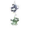


 PDBj
PDBj


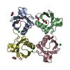
 Assembly


 Mass: 65.409 Da / Num. of mol.: 7 / Source method: obtained synthetically / Formula: Zn
 Sample preparation
 / Beamline: ID14-2 / Wavelength: 0.933
  Processing