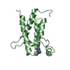
 Movie
Movie Controller


+ Open data

- Basic information
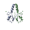
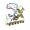
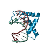
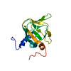
| Entry | Database: PDB / ID: 5o6t | |||||||||
|---|---|---|---|---|---|---|---|---|---|---|
| Title | BIRC4 RING in complex with dimeric ubiquitin variant | |||||||||
 Components Components |
| |||||||||
 Keywords Keywords | LIGASE / E3 ring ligase / ubiquitin variant | |||||||||
| Function / homology |  Function and homology information Function and homology informationregulation of apoptosis involved in tissue homeostasis / positive regulation of protein linear polyubiquitination / regulation of BMP signaling pathway / nucleotide-binding oligomerization domain containing 1 signaling pathway / copper ion homeostasis / regulation of nucleotide-binding domain, leucine rich repeat containing receptor signaling pathway / nucleotide-binding oligomerization domain containing 2 signaling pathway / SMAC, XIAP-regulated apoptotic response / cysteine-type endopeptidase inhibitor activity involved in apoptotic process / Activation of caspases through apoptosome-mediated cleavage ...regulation of apoptosis involved in tissue homeostasis / positive regulation of protein linear polyubiquitination / regulation of BMP signaling pathway / nucleotide-binding oligomerization domain containing 1 signaling pathway / copper ion homeostasis / regulation of nucleotide-binding domain, leucine rich repeat containing receptor signaling pathway / nucleotide-binding oligomerization domain containing 2 signaling pathway / SMAC, XIAP-regulated apoptotic response / cysteine-type endopeptidase inhibitor activity involved in apoptotic process / Activation of caspases through apoptosome-mediated cleavage / SMAC (DIABLO) binds to IAPs / SMAC(DIABLO)-mediated dissociation of IAP:caspase complexes / Regulation of the apoptosome activity / hypothalamus gonadotrophin-releasing hormone neuron development / female meiosis I / TNFR1-induced proapoptotic signaling / positive regulation of protein monoubiquitination / fat pad development / seminiferous tubule development / RIPK1-mediated regulated necrosis / mitochondrion transport along microtubule / regulation of innate immune response / female gonad development / male meiosis I / positive regulation of intrinsic apoptotic signaling pathway by p53 class mediator / negative regulation of tumor necrosis factor-mediated signaling pathway / cysteine-type endopeptidase inhibitor activity / protein K63-linked ubiquitination / energy homeostasis / positive regulation of type I interferon production / neuron projection morphogenesis / Maturation of protein E / Maturation of protein E / ER Quality Control Compartment (ERQC) / Myoclonic epilepsy of Lafora / FLT3 signaling by CBL mutants / IRAK2 mediated activation of TAK1 complex / Alpha-protein kinase 1 signaling pathway / Glycogen synthesis / IRAK1 recruits IKK complex / IRAK1 recruits IKK complex upon TLR7/8 or 9 stimulation / Prevention of phagosomal-lysosomal fusion / Endosomal Sorting Complex Required For Transport (ESCRT) / Membrane binding and targetting of GAG proteins / Regulation of TBK1, IKKε (IKBKE)-mediated activation of IRF3, IRF7 / Negative regulation of FLT3 / PTK6 Regulates RTKs and Their Effectors AKT1 and DOK1 / Regulation of TBK1, IKKε-mediated activation of IRF3, IRF7 upon TLR3 ligation / IRAK2 mediated activation of TAK1 complex upon TLR7/8 or 9 stimulation / Constitutive Signaling by NOTCH1 HD Domain Mutants / NOTCH2 Activation and Transmission of Signal to the Nucleus / TICAM1,TRAF6-dependent induction of TAK1 complex / regulation of proteasomal protein catabolic process / TICAM1-dependent activation of IRF3/IRF7 / APC/C:Cdc20 mediated degradation of Cyclin B / regulation of neuron apoptotic process / Downregulation of ERBB4 signaling / APC-Cdc20 mediated degradation of Nek2A / Regulation of FZD by ubiquitination / p75NTR recruits signalling complexes / InlA-mediated entry of Listeria monocytogenes into host cells / TRAF6 mediated IRF7 activation in TLR7/8 or 9 signaling / NF-kB is activated and signals survival / TRAF6-mediated induction of TAK1 complex within TLR4 complex / Regulation of pyruvate metabolism / positive regulation of protein ubiquitination / Pexophagy / Downregulation of ERBB2:ERBB3 signaling / Regulation of innate immune responses to cytosolic DNA / NRIF signals cell death from the nucleus / Regulation of PTEN localization / regulation of mitochondrial membrane potential / VLDLR internalisation and degradation / Activated NOTCH1 Transmits Signal to the Nucleus / Synthesis of active ubiquitin: roles of E1 and E2 enzymes / Translesion synthesis by REV1 / TICAM1, RIP1-mediated IKK complex recruitment / Regulation of BACH1 activity / Translesion synthesis by POLK / JNK (c-Jun kinases) phosphorylation and activation mediated by activated human TAK1 / InlB-mediated entry of Listeria monocytogenes into host cell / protein serine/threonine kinase binding / MAP3K8 (TPL2)-dependent MAPK1/3 activation / Activation of IRF3, IRF7 mediated by TBK1, IKKε (IKBKE) / Downregulation of TGF-beta receptor signaling / Translesion synthesis by POLI / Josephin domain DUBs / Gap-filling DNA repair synthesis and ligation in GG-NER / IKK complex recruitment mediated by RIP1 / PINK1-PRKN Mediated Mitophagy / TGF-beta receptor signaling in EMT (epithelial to mesenchymal transition) / TNFR1-induced NF-kappa-B signaling pathway / Regulation of activated PAK-2p34 by proteasome mediated degradation / TCF dependent signaling in response to WNT / Regulation of NF-kappa B signaling / activated TAK1 mediates p38 MAPK activation / Autodegradation of Cdh1 by Cdh1:APC/C / APC/C:Cdc20 mediated degradation of Securin / NOTCH3 Activation and Transmission of Signal to the Nucleus / Regulation of signaling by CBL Similarity search - Function | |||||||||
| Biological species |  Homo sapiens (human) Homo sapiens (human) | |||||||||
| Method |  X-RAY DIFFRACTION / SYNCHROTRON / MOLECULAR REPLACEMENT / Resolution: 1.57 Å X-RAY DIFFRACTION / SYNCHROTRON / MOLECULAR REPLACEMENT / Resolution: 1.57 Å | |||||||||
 Authors Authors | Gabrielsen, M. / Buetow, L. / Huang, D.T. | |||||||||
| Funding support |  United Kingdom, 2items United Kingdom, 2items
| |||||||||
 Citation Citation | Journal: Mol. Cell / Year: 2017 Title: A General Strategy for Discovery of Inhibitors and Activators of RING and U-box E3 Ligases with Ubiquitin Variants. Authors: Gabrielsen, M. / Buetow, L. / Nakasone, M.A. / Ahmed, S.F. / Sibbet, G.J. / Smith, B.O. / Zhang, W. / Sidhu, S.S. / Huang, D.T. | |||||||||
| History |
|
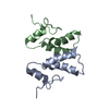
- Structure visualization
| Structure viewer | Molecule: MolmilJmol/JSmol |
|---|
- Downloads & links
-Download
| PDBx/mmCIF format | 5o6t.cif.gz | 241.6 KB | Display | PDBx/mmCIF format |
|---|---|---|---|---|
| PDB format | pdb5o6t.ent.gz | 197.5 KB | Display | PDB format |
| PDBx/mmJSON format | 5o6t.json.gz | Tree view | PDBx/mmJSON format | |
| Others |  Other downloads Other downloads |
-Validation report
| Arichive directory | https://data.pdbj.org/pub/pdb/validation_reports/o6/5o6tftp://data.pdbj.org/pub/pdb/validation_reports/o6/5o6t | HTTPS FTP |
|---|
-Related structure data
| Related structure data | 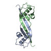 5o6sC 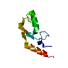 5o75C 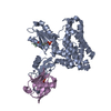 5o76C  1ubqS  3eb5S S: Starting model for refinement C: citing same article ( |
|---|---|
| Similar structure data |


-Links
 PDBj
PDBj


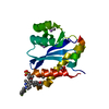
- Assembly
| Deposited unit | 
| ||||||||
|---|---|---|---|---|---|---|---|---|---|
| 1 | 
| ||||||||
| 2 | 
| ||||||||
| Unit cell |
|
-Components
| #1: Protein | Mass: 7569.029 Da / Num. of mol.: 2 Source method: isolated from a genetically manipulated source Source: (gene. exp.) Homo sapiens (human) / Gene: XIAP, API3, BIRC4, IAP3 / Plasmid: pGEX4T.1 / Production host:  References: UniProt: P98170, RING-type E3 ubiquitin transferase #2: Protein | Mass: 9041.301 Da / Num. of mol.: 2 Source method: isolated from a genetically manipulated source Details: A variant of ubiquitin identified from a phage-display library Source: (gene. exp.) Homo sapiens (human) / Gene: UBB / Production host: #3: Chemical | ChemComp-ZN /   Mass: 65.409 Da / Num. of mol.: 4 / Source method: obtained synthetically / Formula: Zn Mass: 65.409 Da / Num. of mol.: 4 / Source method: obtained synthetically / Formula: Zn#4: Chemical |   Mass: 62.068 Da / Num. of mol.: 2 / Source method: obtained synthetically / Formula: C2H6O2 Mass: 62.068 Da / Num. of mol.: 2 / Source method: obtained synthetically / Formula: C2H6O2#5: Water | ChemComp-HOH / |  Mass: 18.015 Da / Num. of mol.: 218 / Source method: isolated from a natural source / Formula: H2O Mass: 18.015 Da / Num. of mol.: 218 / Source method: isolated from a natural source / Formula: H2O |
|---|
-Experimental details
-Experiment
| Experiment | Method: X-RAY DIFFRACTION / Number of used crystals: 1 |
|---|
- Sample preparation
| Crystal | Density Matthews: 2.06 Å3/Da / Density % sol: 40.41 % / Description: Rectangular cuboids |
|---|---|
| Crystal grow | Temperature: 291 K / Method: vapor diffusion, sitting drop / pH: 7.6 / Details: 0.1 M NaHEPES pH 7.5, 1.4 M tri-Na citrate |
-Data collection
| Diffraction | Mean temperature: 110 K |
|---|---|
| Diffraction source | Source: SYNCHROTRON / Site: Diamond / Beamline: I04 / Wavelength: 0.9795 Å |
| Detector | Type: DECTRIS PILATUS 6M-F / Detector: PIXEL / Date: Apr 28, 2016 |
| Radiation | Protocol: SINGLE WAVELENGTH / Monochromatic (M) / Laue (L): M / Scattering type: x-ray |
| Radiation wavelength | Wavelength: 0.9795 Å / Relative weight: 1 |
| Reflection | Resolution: 1.57→69.97 Å / Num. obs: 39227 / % possible obs: 100 % / Redundancy: 6.4 % / Biso Wilson estimate: 20.57 Å2 / Rmerge(I) obs: 0.074 / Rpim(I) all: 0.042 / Net I/σ(I): 14.9 |
| Reflection shell | Resolution: 1.57→1.577 Å / Redundancy: 6.5 % / Rmerge(I) obs: 0.89 / Mean I/σ(I) obs: 2 / Num. unique obs: 2560 / CC1/2: 0.663 / Rpim(I) all: 0.513 / % possible all: 96.3 |
- Processing
| Software |
| |||||||||||||||||||||||||||||||||||||||||||||||||||||||||||||||||||||||||||||||||||||||||||||||||||||||||||||||||||||||||||||
|---|---|---|---|---|---|---|---|---|---|---|---|---|---|---|---|---|---|---|---|---|---|---|---|---|---|---|---|---|---|---|---|---|---|---|---|---|---|---|---|---|---|---|---|---|---|---|---|---|---|---|---|---|---|---|---|---|---|---|---|---|---|---|---|---|---|---|---|---|---|---|---|---|---|---|---|---|---|---|---|---|---|---|---|---|---|---|---|---|---|---|---|---|---|---|---|---|---|---|---|---|---|---|---|---|---|---|---|---|---|---|---|---|---|---|---|---|---|---|---|---|---|---|---|---|---|---|
| Refinement | Method to determine structure: MOLECULAR REPLACEMENT Starting model: 3EB5, 1UBQ Resolution: 1.57→43.18 Å / Cor.coef. Fo:Fc: 0.964 / Cor.coef. Fo:Fc free: 0.955 / Rfactor Rfree error: 0 / SU R Cruickshank DPI: 0.088 / Cross valid method: THROUGHOUT / σ(F): 0 / SU R Blow DPI: 0.076 / SU Rfree Blow DPI: 0.076 / SU Rfree Cruickshank DPI: 0.073
| |||||||||||||||||||||||||||||||||||||||||||||||||||||||||||||||||||||||||||||||||||||||||||||||||||||||||||||||||||||||||||||
| Displacement parameters | Biso mean: 29.69 Å2
| |||||||||||||||||||||||||||||||||||||||||||||||||||||||||||||||||||||||||||||||||||||||||||||||||||||||||||||||||||||||||||||
| Refine analyze | Luzzati coordinate error obs: 0.17 Å | |||||||||||||||||||||||||||||||||||||||||||||||||||||||||||||||||||||||||||||||||||||||||||||||||||||||||||||||||||||||||||||
| Refinement step | Cycle: 1 / Resolution: 1.57→43.18 Å
| |||||||||||||||||||||||||||||||||||||||||||||||||||||||||||||||||||||||||||||||||||||||||||||||||||||||||||||||||||||||||||||
| Refine LS restraints |
| |||||||||||||||||||||||||||||||||||||||||||||||||||||||||||||||||||||||||||||||||||||||||||||||||||||||||||||||||||||||||||||
| LS refinement shell | Resolution: 1.57→1.61 Å / Rfactor Rfree error: 0 / Total num. of bins used: 20
| |||||||||||||||||||||||||||||||||||||||||||||||||||||||||||||||||||||||||||||||||||||||||||||||||||||||||||||||||||||||||||||
| Refinement TLS params. | Method: refined / Refine-ID: X-RAY DIFFRACTION
| |||||||||||||||||||||||||||||||||||||||||||||||||||||||||||||||||||||||||||||||||||||||||||||||||||||||||||||||||||||||||||||
| Refinement TLS group |
|
