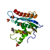
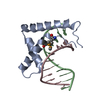
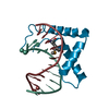
- PDB-6jrp: Crystal structure of CIC-HMG-ETV5-DNA complex -
+
Open data
ID or keywords:
Loading...
-
Basic information
Entry
Database: PDB / ID: 6jrp
Title
Crystal structure of CIC-HMG-ETV5-DNA complex
Components
DNA (5'-D(*AP*TP*GP*AP*AP*TP*GP*AP*AP*AP*A)-3')
DNA (5'-D(*TP*TP*TP*TP*CP*AP*TP*TP*CP*AP*T)-3')
Protein capicua homolog
Keywords
TRANSCRIPTION / repressor / protein-DNA complex
Function / homology
Function and homology information
lung alveolus development / social behavior / learning / RNA polymerase II transcription regulatory region sequence-specific DNA binding / brain development / memory / transcription by RNA polymerase II / DNA-binding transcription factor activity, RNA polymerase II-specific / negative regulation of DNA-templated transcription / chromatin binding ...lung alveolus development / social behavior / learning / RNA polymerase II transcription regulatory region sequence-specific DNA binding / brain development / memory / transcription by RNA polymerase II / DNA-binding transcription factor activity, RNA polymerase II-specific / negative regulation of DNA-templated transcription / chromatin binding / regulation of transcription by RNA polymerase II / chromatin / negative regulation of transcription by RNA polymerase II / protein-containing complex / nucleoplasm / nucleus Similarity search - Function
Domain of unknown function DUF4819 / : / : / Protein capicua homolog-like domain / Protein capicua homolog-like, C-terminal tri-helical domain / : / High mobility group box domain / DNA Binding (I), subunit A / HMG (high mobility group) box / HMG boxes A and B DNA-binding domains profile. ...Domain of unknown function DUF4819 / : / : / Protein capicua homolog-like domain / Protein capicua homolog-like, C-terminal tri-helical domain / : / High mobility group box domain / DNA Binding (I), subunit A / HMG (high mobility group) box / HMG boxes A and B DNA-binding domains profile. / high mobility group / High mobility group box domain / High mobility group box domain superfamily / Orthogonal Bundle / Mainly Alpha Similarity search - Domain/homology
Journal: Febs J. / Year: 2019 Title: The crystal structure of Capicua HMG-box domain complexed with the ETV5-DNA and its implications for Capicua-mediated cancers. Authors: Lee, H. / Song, J.J.
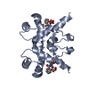
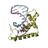
A: Protein capicua homolog B: DNA (5'-D(*AP*TP*GP*AP*AP*TP*GP*AP*AP*AP*A)-3') C: DNA (5'-D(*TP*TP*TP*TP*CP*AP*TP*TP*CP*AP*T)-3') D: Protein capicua homolog E: DNA (5'-D(*AP*TP*GP*AP*AP*TP*GP*AP*AP*AP*A)-3') F: DNA (5'-D(*TP*TP*TP*TP*CP*AP*TP*TP*CP*AP*T)-3') G: Protein capicua homolog H: DNA (5'-D(*AP*TP*GP*AP*AP*TP*GP*AP*AP*AP*A)-3') I: DNA (5'-D(*TP*TP*TP*TP*CP*AP*TP*TP*CP*AP*T)-3') J: Protein capicua homolog K: DNA (5'-D(*AP*TP*GP*AP*AP*TP*GP*AP*AP*AP*A)-3') L: DNA (5'-D(*TP*TP*TP*TP*CP*AP*TP*TP*CP*AP*T)-3')
In the structure databanks used in Yorodumi, some data are registered as the other names, "COVID-19 virus" and "2019-nCoV". Here are the details of the virus and the list of structure data.
Jan 31, 2019. EMDB accession codes are about to change! (news from PDBe EMDB page)
EMDB accession codes are about to change! (news from PDBe EMDB page)
The allocation of 4 digits for EMDB accession codes will soon come to an end. Whilst these codes will remain in use, new EMDB accession codes will include an additional digit and will expand incrementally as the available range of codes is exhausted. The current 4-digit format prefixed with “EMD-” (i.e. EMD-XXXX) will advance to a 5-digit format (i.e. EMD-XXXXX), and so on. It is currently estimated that the 4-digit codes will be depleted around Spring 2019, at which point the 5-digit format will come into force.
The EM Navigator/Yorodumi systems omit the EMD- prefix.
Related info.:Q: What is EMD? / ID/Accession-code notation in Yorodumi/EM Navigator
Yorodumi is a browser for structure data from EMDB, PDB, SASBDB, etc.
This page is also the successor to EM Navigator detail page, and also detail information page/front-end page for Omokage search.
The word "yorodu" (or yorozu) is an old Japanese word meaning "ten thousand". "mi" (miru) is to see.
Related info.:EMDB / PDB / SASBDB / Comparison of 3 databanks / Yorodumi Search / Aug 31, 2016. New EM Navigator & Yorodumi / Yorodumi Papers / Jmol/JSmol / Function and homology information / Changes in new EM Navigator and Yorodumi
 Movie
Movie Controller


 Open data
 Basic information
 Components
 Keywords
 Function and homology information
 Homo sapiens (human)
 X-RAY DIFFRACTION /
  Authors
 Korea, Republic Of, 2items
  Citation
 Structure visualization
 Downloads & links
 Other downloads
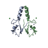


 PDBj
PDBj


 Assembly


 Sample preparation
 / Beamline: AR-NE3A / Wavelength: 0.9778, 0.9785
 Processing