 Movie
Movie Controller

[English] 日本語
 Yorodumi
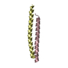
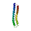
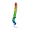
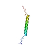
- PDB-2x9c: Crystal structure of a soluble PrgI mutant from Salmonella Typhimurium -
+ Open data

- Basic information
| Entry | Database: PDB / ID: 2x9c | ||||||
|---|---|---|---|---|---|---|---|
| Title | Crystal structure of a soluble PrgI mutant from Salmonella Typhimurium | ||||||
 Components Components | PROTEIN PRGI | ||||||
 Keywords Keywords | PROTEIN TRANSPORT / NEEDLE PROTOMER / BACTERIAL PATHOGENESIS | ||||||
| Function / homology |  Function and homology information Function and homology informationtype III protein secretion system complex / protein secretion by the type III secretion system / cell surface / extracellular region / identical protein binding Similarity search - Function | ||||||
| Biological species |  SALMONELLA TYPHIMURIUM (bacteria) SALMONELLA TYPHIMURIUM (bacteria) | ||||||
| Method |  X-RAY DIFFRACTION / SYNCHROTRON / MOLECULAR REPLACEMENT / Resolution: 2.45 Å X-RAY DIFFRACTION / SYNCHROTRON / MOLECULAR REPLACEMENT / Resolution: 2.45 Å | ||||||
 Authors Authors | Poyraz, O. / Schmidt, H. / Seidel, K. / Delissen, F. / Ader, C. / Tenenboim, H. / Goosmann, C. / Laube, B. / Thuenemann, A.F. / Zychlinsky, A. ...Poyraz, O. / Schmidt, H. / Seidel, K. / Delissen, F. / Ader, C. / Tenenboim, H. / Goosmann, C. / Laube, B. / Thuenemann, A.F. / Zychlinsky, A. / Baldus, M. / Lange, A. / Griesinger, C. / Kolbe, M. | ||||||
 Citation Citation | Journal: Nat.Struct.Mol.Biol. / Year: 2010 Title: Protein Refolding is Required for Assembly of the Type Three Secretion Needle Authors: Poyraz, O. / Schmidt, H. / Seidel, K. / Delissen, F. / Ader, C. / Tenenboim, H. / Goosmann, C. / Laube, B. / Thuenemann, A.F. / Zychlinsky, A. / Baldus, M. / Lange, A. / Griesinger, C. / Kolbe, M. | ||||||
| History |
|
- Structure visualization
| Structure viewer | Molecule: MolmilJmol/JSmol |
|---|
- Downloads & links
-Download
| PDBx/mmCIF format | 2x9c.cif.gz | 36.7 KB | Display | PDBx/mmCIF format |
|---|---|---|---|---|
| PDB format | pdb2x9c.ent.gz | 24.9 KB | Display | PDB format |
| PDBx/mmJSON format | 2x9c.json.gz | Tree view | PDBx/mmJSON format | |
| Others |  Other downloads Other downloads |
-Validation report
| Arichive directory | https://data.pdbj.org/pub/pdb/validation_reports/x9/2x9cftp://data.pdbj.org/pub/pdb/validation_reports/x9/2x9c | HTTPS FTP |
|---|
-Related structure data
| Related structure data |  2kv7C 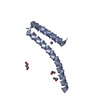 2ca5S C: citing same article ( S: Starting model for refinement |
|---|---|
| Similar structure data |


-Links
 PDBj
PDBj- Assembly
| Deposited unit | 
| ||||||||
|---|---|---|---|---|---|---|---|---|---|
| 1 | 
| ||||||||
| 2 | 
| ||||||||
| Unit cell |
| ||||||||
| Noncrystallographic symmetry (NCS) | NCS oper: (Code: given Matrix: (0.9934, -0.09595, 0.06291), Vector: |
-Components
| #1: Protein | Mass: 9091.037 Da / Num. of mol.: 2 / Mutation: YES Source method: isolated from a genetically manipulated source Source: (gene. exp.) SALMONELLA TYPHIMURIUM (bacteria) / Strain: SL1344 / Plasmid: PET-28A / Production host: #2: Water | ChemComp-HOH / |  Mass: 18.015 Da / Num. of mol.: 9 / Source method: isolated from a natural source / Formula: H2O Mass: 18.015 Da / Num. of mol.: 9 / Source method: isolated from a natural source / Formula: H2OCompound details | ENGINEERED RESIDUE IN CHAIN A, VAL 65 TO ALA ENGINEERED RESIDUE IN CHAIN A, VAL 67 TO ALA ...ENGINEERED | |
|---|
-Experimental details
-Experiment
| Experiment | Method: X-RAY DIFFRACTION / Number of used crystals: 1 |
|---|
- Sample preparation
| Crystal | Density Matthews: 3.45 Å3/Da / Density % sol: 64.4 % Description: DATA DETWINNED USING CNS WITH TWIN FRACTION 0.18 AND TWIN OPERATOR K,H,-L |
|---|---|
| Crystal grow | Method: vapor diffusion, hanging drop Details: RESERVOIR SOLUTION 0.15 MM NAH2PO4. SAMPLE BUFFER 20 MM HEPES (PH 7.5) 50 MM NACL. HANGING DROP WITH 1 UL SAMPLE AND 1 UL RESERVOIR SOLUTION. |
-Data collection
| Diffraction | Mean temperature: 100 K |
|---|---|
| Diffraction source | Source: SYNCHROTRON / Site: ESRF  / Beamline: ID23-1 / Wavelength: 0.97625 / Beamline: ID23-1 / Wavelength: 0.97625 |
| Detector | Type: ADSC QUANTUM 315r / Detector: CCD / Date: May 13, 2007 / Details: TOROIDAL MIRROR |
| Radiation | Monochromator: SI(111) CHANNEL-CUT / Protocol: SINGLE WAVELENGTH / Monochromatic (M) / Laue (L): M / Scattering type: x-ray |
| Radiation wavelength | Wavelength: 0.97625 Å / Relative weight: 1 |
| Reflection | Resolution: 2.25→50 Å / Num. obs: 11435 / % possible obs: 94.9 % / Observed criterion σ(I): -3 / Redundancy: 7.5 % / Biso Wilson estimate: 63.9 Å2 / Rmerge(I) obs: 0.11 / Net I/σ(I): 9.83 |
| Reflection shell | Resolution: 2.25→2.31 Å / Redundancy: 7.6 % / Rmerge(I) obs: 0.92 / Mean I/σ(I) obs: 2.11 / % possible all: 96.8 |
- Processing
| Software |
| ||||||||||||||||||||||||||||||||||||||||||||||||||||||||||||||||||||||||||||||||
|---|---|---|---|---|---|---|---|---|---|---|---|---|---|---|---|---|---|---|---|---|---|---|---|---|---|---|---|---|---|---|---|---|---|---|---|---|---|---|---|---|---|---|---|---|---|---|---|---|---|---|---|---|---|---|---|---|---|---|---|---|---|---|---|---|---|---|---|---|---|---|---|---|---|---|---|---|---|---|---|---|---|
| Refinement | Method to determine structure: MOLECULAR REPLACEMENT Starting model: PDB ENTRY 2CA5 Resolution: 2.45→38.13 Å / Rfactor Rfree error: 0.012 / Data cutoff high absF: 2660057.81 / Data cutoff low absF: 0 / Isotropic thermal model: RESTRAINED / Cross valid method: THROUGHOUT / σ(F): 0 / Stereochemistry target values: MAXIMUM LIKELIHOOD Details: CHAIN A RESIDUES 1-18 AND 80 ARE DISORDERED. CHAIN B RESIDUES 1-17 ARE DISORDERED. N-TERMINAL RESIDUES GLY-SER-HIS REMAINING FROM THROMBIN CLEAVAGE SITE ARE DISORDERED IN CHAINS A AND B. THE ...Details: CHAIN A RESIDUES 1-18 AND 80 ARE DISORDERED. CHAIN B RESIDUES 1-17 ARE DISORDERED. N-TERMINAL RESIDUES GLY-SER-HIS REMAINING FROM THROMBIN CLEAVAGE SITE ARE DISORDERED IN CHAINS A AND B. THE STRUCTURE WAS REFINED AT LOWER RESOLUTION (2.45 A) THAN THE COLLECTED DATASET (2.25 A) BECAUSE OF THE POOR MERGING STATISTICS AT HIGH RESOLUTION.
| ||||||||||||||||||||||||||||||||||||||||||||||||||||||||||||||||||||||||||||||||
| Solvent computation | Solvent model: FLAT MODEL / Bsol: 79.6715 Å2 / ksol: 0.4 e/Å3 | ||||||||||||||||||||||||||||||||||||||||||||||||||||||||||||||||||||||||||||||||
| Displacement parameters | Biso mean: 81.6 Å2
| ||||||||||||||||||||||||||||||||||||||||||||||||||||||||||||||||||||||||||||||||
| Refine analyze |
| ||||||||||||||||||||||||||||||||||||||||||||||||||||||||||||||||||||||||||||||||
| Refinement step | Cycle: LAST / Resolution: 2.45→38.13 Å
| ||||||||||||||||||||||||||||||||||||||||||||||||||||||||||||||||||||||||||||||||
| Refine LS restraints |
| ||||||||||||||||||||||||||||||||||||||||||||||||||||||||||||||||||||||||||||||||
| Refine LS restraints NCS | NCS model details: NONE | ||||||||||||||||||||||||||||||||||||||||||||||||||||||||||||||||||||||||||||||||
| LS refinement shell | Resolution: 2.45→2.6 Å / Rfactor Rfree error: 0.071 / Total num. of bins used: 6
| ||||||||||||||||||||||||||||||||||||||||||||||||||||||||||||||||||||||||||||||||
| Xplor file |
|
