 Movie
Movie Controller


+ Open data

- Basic information
| Entry | Database: PDB / ID: 2we5 | ||||||
|---|---|---|---|---|---|---|---|
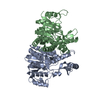
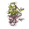
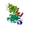
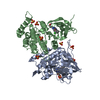
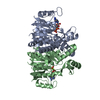
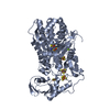
| Title | Carbamate kinase from Enterococcus faecalis bound to MgADP | ||||||
 Components Components | CARBAMATE KINASE 1 | ||||||
 Keywords Keywords | TRANSFERASE / ARGININE CATABOLISM / ARGININE METABOLISM / ATP SYNTHESYS / KINASE / OPEN ALPHA/BETA SHEET / PHOSPHOTRANSFERASE | ||||||
| Function / homology |  Function and homology information Function and homology informationcarbamate kinase / carbamate kinase activity / : / ATP binding / cytosol Similarity search - Function | ||||||
| Biological species |   ENTEROCOCCUS FAECALIS (bacteria) ENTEROCOCCUS FAECALIS (bacteria) | ||||||
| Method |  X-RAY DIFFRACTION / SYNCHROTRON / MOLECULAR REPLACEMENT / Resolution: 1.39 Å X-RAY DIFFRACTION / SYNCHROTRON / MOLECULAR REPLACEMENT / Resolution: 1.39 Å | ||||||
 Authors Authors | Ramon-Maiques, S. / Marina, A. / Rubio, V. | ||||||
 Citation Citation | Journal: J.Mol.Biol. / Year: 2010 Title: Substrate Binding and Catalysis in Carbamate Kinase Ascertained by Crystallographic and Site- Directed Mutagenesis Studies. Movements and Significance of a Unique Globular Subdomain of This ...Title: Substrate Binding and Catalysis in Carbamate Kinase Ascertained by Crystallographic and Site- Directed Mutagenesis Studies. Movements and Significance of a Unique Globular Subdomain of This Key Enzyme for Fermentative ATP Production in Bacteria. Authors: Ramon-Maiques, S. / Marina, A. / Guinot, A. / Gil-Ortiz, F. / Uriarte, M. / Fita, I. / Rubio, V. #1: Journal: Protein Sci. / Year: 1999Title: Carbamate Kinase: New Structural Machinery for Making Carbamoyl Phosphate, the Common Precursor of Pyrimidines and Arginine. Authors: Marina, A. / Alzari, P.M. / Bravo, J. / Uriarte, M. / Barcelona, B. / Fita, I. / Rubio, V. #2: Journal: J.Mol.Biol. / Year: 2000Title: The 1.5 A Resolution Crystal Structure of the Carbamate Kinase-Like Carbamoyl Phosphate Synthetase from the Hyperthermophilic Archaeon Pyrococcus Furiosus, Bound to Adp, Confirms that This ...Title: The 1.5 A Resolution Crystal Structure of the Carbamate Kinase-Like Carbamoyl Phosphate Synthetase from the Hyperthermophilic Archaeon Pyrococcus Furiosus, Bound to Adp, Confirms that This Thermostable Enzyme is a Carbamate Kinase, and Provides Insight Into Substrate Binding and Stability in Carbamate Kinases. Authors: Ramon-Maiques, S. / Marina, A. / Uriarte, M. / Fita, I. / Rubio, V. | ||||||
| History |
|
- Structure visualization
| Structure viewer | Molecule: MolmilJmol/JSmol |
|---|
- Downloads & links
-Download
| PDBx/mmCIF format | 2we5.cif.gz | 205.8 KB | Display | PDBx/mmCIF format |
|---|---|---|---|---|
| PDB format | pdb2we5.ent.gz | 164.7 KB | Display | PDB format |
| PDBx/mmJSON format | 2we5.json.gz | Tree view | PDBx/mmJSON format | |
| Others |  Other downloads Other downloads |
-Validation report
| Arichive directory | https://data.pdbj.org/pub/pdb/validation_reports/we/2we5ftp://data.pdbj.org/pub/pdb/validation_reports/we/2we5 | HTTPS FTP |
|---|
-Related structure data
| Related structure data | 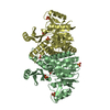 2we4C 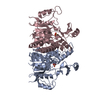 1b7bS S: Starting model for refinement C: citing same article ( |
|---|---|
| Similar structure data |

-Links
 PDBj
PDBj- Assembly
| Deposited unit | 
| ||||||||
|---|---|---|---|---|---|---|---|---|---|
| 1 | 
| ||||||||
| 2 | 
| ||||||||
| Unit cell |
|
-Components
| #1: Protein | Mass: 32958.578 Da / Num. of mol.: 3 Source method: isolated from a genetically manipulated source Source: (gene. exp.) ENTEROCOCCUS FAECALIS (bacteria) / Plasmid: PET15B / Production host: #2: Chemical |   Mass: 427.201 Da / Num. of mol.: 3 / Source method: obtained synthetically / Formula: C10H15N5O10P2 / Comment: ADP, energy-carrying molecule*YM Mass: 427.201 Da / Num. of mol.: 3 / Source method: obtained synthetically / Formula: C10H15N5O10P2 / Comment: ADP, energy-carrying molecule*YM#3: Chemical |   Mass: 24.305 Da / Num. of mol.: 3 / Source method: obtained synthetically / Formula: Mg Mass: 24.305 Da / Num. of mol.: 3 / Source method: obtained synthetically / Formula: Mg#4: Chemical | ChemComp-ACT / |   Mass: 59.044 Da / Num. of mol.: 1 / Source method: obtained synthetically / Formula: C2H3O2 Mass: 59.044 Da / Num. of mol.: 1 / Source method: obtained synthetically / Formula: C2H3O2#5: Water | ChemComp-HOH / |  Mass: 18.015 Da / Num. of mol.: 840 / Source method: isolated from a natural source / Formula: H2O Mass: 18.015 Da / Num. of mol.: 840 / Source method: isolated from a natural source / Formula: H2O |
|---|
-Experimental details
-Experiment
| Experiment | Method: X-RAY DIFFRACTION / Number of used crystals: 1 |
|---|
- Sample preparation
| Crystal | Density Matthews: 2.25 Å3/Da / Density % sol: 45 % / Description: NONE |
|---|---|
| Crystal grow | Temperature: 277 K / Method: vapor diffusion, hanging drop Details: CRYSTALS WERE OBTAINED AT 4 DEGREES USING THE HANGING DROP VAPOUR DIFFUSION METHOD BY MIXING 1.5 MIRCROLITERS OF A 10 MG/ML PROTEIN SOLUTION IN 5 MM SODIUM CACODYLATE PH 6.5, 2 MM ADP, 5 MM ...Details: CRYSTALS WERE OBTAINED AT 4 DEGREES USING THE HANGING DROP VAPOUR DIFFUSION METHOD BY MIXING 1.5 MIRCROLITERS OF A 10 MG/ML PROTEIN SOLUTION IN 5 MM SODIUM CACODYLATE PH 6.5, 2 MM ADP, 5 MM MGCL2 AND 50 MM SODIUM PHOPHONOACETATE, AND 1.5 MICROLITERS OF THE RESERVOIR BUFFER CONTAINING 13-16% PEG 8000, 130-160 MM MAGNESIUM ACETATE AND 0.1 M SODIUM CACODYLATE PH 6.5 |
-Data collection
| Diffraction | Mean temperature: 100 K |
|---|---|
| Diffraction source | Source: SYNCHROTRON / Site: EMBL/DESY, HAMBURG  / Beamline: X11 / Wavelength: 0.9067 / Beamline: X11 / Wavelength: 0.9067 |
| Detector | Type: MARRESEARCH / Detector: IMAGE PLATE / Date: Jul 1, 1998 |
| Radiation | Protocol: SINGLE WAVELENGTH / Monochromatic (M) / Laue (L): M / Scattering type: x-ray |
| Radiation wavelength | Wavelength: 0.9067 Å / Relative weight: 1 |
| Reflection | Resolution: 1.39→50 Å / Num. obs: 172839 / % possible obs: 90.4 % / Observed criterion σ(I): 0 / Redundancy: 3.3 % / Biso Wilson estimate: 20 Å2 / Rmerge(I) obs: 0.05 / Net I/σ(I): 7 |
| Reflection shell | Resolution: 1.39→1.47 Å / Redundancy: 2.9 % / Rmerge(I) obs: 0.33 / Mean I/σ(I) obs: 2.2 / % possible all: 87.1 |
- Processing
| Software |
| ||||||||||||||||||||||||||||||||||||||||||||||||||||||||||||
|---|---|---|---|---|---|---|---|---|---|---|---|---|---|---|---|---|---|---|---|---|---|---|---|---|---|---|---|---|---|---|---|---|---|---|---|---|---|---|---|---|---|---|---|---|---|---|---|---|---|---|---|---|---|---|---|---|---|---|---|---|---|
| Refinement | Method to determine structure: MOLECULAR REPLACEMENT Starting model: PDB ENTRY 1B7B Resolution: 1.39→20 Å / Data cutoff high absF: 10000 / Cross valid method: THROUGHOUT / σ(F): 0
| ||||||||||||||||||||||||||||||||||||||||||||||||||||||||||||
| Solvent computation | Bsol: 46.1408 Å2 / ksol: 0.342447 e/Å3 | ||||||||||||||||||||||||||||||||||||||||||||||||||||||||||||
| Displacement parameters | Biso mean: 24.7 Å2
| ||||||||||||||||||||||||||||||||||||||||||||||||||||||||||||
| Refinement step | Cycle: LAST / Resolution: 1.39→20 Å
| ||||||||||||||||||||||||||||||||||||||||||||||||||||||||||||
| Refine LS restraints |
| ||||||||||||||||||||||||||||||||||||||||||||||||||||||||||||
| Xplor file |
|
