 Movie
Movie Controller

[English] 日本語
 Yorodumi
- PDB-1ais: TATA-BINDING PROTEIN/TRANSCRIPTION FACTOR (II)B/TATA-BOX COMPLEX ... -
+ Open data

- Basic information
| Entry | Database: PDB / ID: 1ais | ||||||
|---|---|---|---|---|---|---|---|
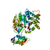
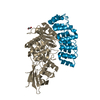
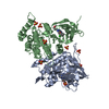
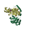
| Title | TATA-BINDING PROTEIN/TRANSCRIPTION FACTOR (II)B/TATA-BOX COMPLEX FROM PYROCOCCUS WOESEI | ||||||
 Components Components |
| ||||||
 Keywords Keywords | TRANSCRIPTION/DNA / TRANSCRIPTION / HYPERTHERMOPHILE / RIBOSOME BINDING / COMPLEX (RIBOSOME BINDING- DNA) / TRANSCRIPTION-DNA COMPLEX | ||||||
| Function / homology |  Function and homology information Function and homology informationtranscription preinitiation complex assembly / transcription preinitiation complex / TBP-class protein binding / DNA-templated transcription initiation / DNA-binding transcription factor activity / DNA binding / zinc ion binding Similarity search - Function | ||||||
| Biological species |   Pyrococcus woesei (archaea) Pyrococcus woesei (archaea) | ||||||
| Method |  X-RAY DIFFRACTION / SYNCHROTRON / MIR / Resolution: 2.1 Å X-RAY DIFFRACTION / SYNCHROTRON / MIR / Resolution: 2.1 Å | ||||||
 Authors Authors | Kosa, P.F. / Ghosh, G. / Dedecker, B.S. / Sigler, P.B. | ||||||
 Citation Citation | Journal: Proc.Natl.Acad.Sci.USA / Year: 1997 Title: The 2.1-A crystal structure of an archaeal preinitiation complex: TATA-box-binding protein/transcription factor (II)B core/TATA-box. Authors: Kosa, P.F. / Ghosh, G. / DeDecker, B.S. / Sigler, P.B. | ||||||
| History |
|
- Structure visualization
| Structure viewer | Molecule: MolmilJmol/JSmol |
|---|
- Downloads & links
-Download
| PDBx/mmCIF format | 1ais.cif.gz | 114.5 KB | Display | PDBx/mmCIF format |
|---|---|---|---|---|
| PDB format | pdb1ais.ent.gz | 84 KB | Display | PDB format |
| PDBx/mmJSON format | 1ais.json.gz | Tree view | PDBx/mmJSON format | |
| Others |  Other downloads Other downloads |
-Validation report
| Arichive directory | https://data.pdbj.org/pub/pdb/validation_reports/ai/1aisftp://data.pdbj.org/pub/pdb/validation_reports/ai/1ais | HTTPS FTP |
|---|
-Related structure data
| Similar structure data |
|---|


-Links
 PDBj
PDBj


- Assembly
| Deposited unit | 
| ||||||||
|---|---|---|---|---|---|---|---|---|---|
| 1 |
| ||||||||
| Unit cell |
|
-Components
| #1: DNA chain | Mass: 5384.125 Da / Num. of mol.: 1 / Source method: obtained synthetically |
|---|---|
| #2: DNA chain | Mass: 5249.446 Da / Num. of mol.: 1 / Source method: obtained synthetically |
| #3: Protein | Mass: 20279.707 Da / Num. of mol.: 1 / Fragment: RESIDUES 1 - 181 Source method: isolated from a genetically manipulated source Source: (gene. exp.) Pyrococcus woesei (archaea) / Plasmid: PET11A / Species (production host): Escherichia coli / Production host:  |
| #4: Protein | Mass: 22590.379 Da / Num. of mol.: 1 / Fragment: C TERMINAL DOMAIN Source method: isolated from a genetically manipulated source Source: (gene. exp.) Pyrococcus woesei (archaea) / Plasmid: PET11A / Species (production host): Escherichia coli / Production host: |
| #5: Water | ChemComp-HOH /  Mass: 18.015 Da / Num. of mol.: 288 / Source method: isolated from a natural source / Formula: H2O Mass: 18.015 Da / Num. of mol.: 288 / Source method: isolated from a natural source / Formula: H2O |
| Has protein modification | Y |
-Experimental details
-Experiment
| Experiment | Method: X-RAY DIFFRACTION / Number of used crystals: 1 |
|---|
- Sample preparation
| Crystal | Density Matthews: 3.34 Å3/Da / Density % sol: 64 % | ||||||||||||||||||||||||||||||||||||||||
|---|---|---|---|---|---|---|---|---|---|---|---|---|---|---|---|---|---|---|---|---|---|---|---|---|---|---|---|---|---|---|---|---|---|---|---|---|---|---|---|---|---|
| Crystal grow | Method: vapor diffusion, hanging drop / pH: 7.4 Details: HANGING DROP METHOD 8% PEG 8000, 200 MM POTASSIUM PHOSPHATE PH 7.4., vapor diffusion - hanging drop | ||||||||||||||||||||||||||||||||||||||||
| Components of the solutions |
| ||||||||||||||||||||||||||||||||||||||||
| Crystal | *PLUS Density % sol: 64 % | ||||||||||||||||||||||||||||||||||||||||
| Crystal grow | *PLUS Temperature: 18 ℃ | ||||||||||||||||||||||||||||||||||||||||
| Components of the solutions | *PLUS
|
-Data collection
| Diffraction | Mean temperature: 100 K |
|---|---|
| Diffraction source | Source: SYNCHROTRON / Site: CHESS  / Beamline: A1 / Beamline: A1 |
| Detector | Detector: CCD / Date: Jan 1, 1996 / Details: MIRRORS |
| Radiation | Protocol: SINGLE WAVELENGTH / Monochromatic (M) / Laue (L): M / Scattering type: x-ray |
| Radiation wavelength | Relative weight: 1 |
| Reflection | Resolution: 2.1→20 Å / Num. obs: 14445 / % possible obs: 92 % / Observed criterion σ(F): 3 / Observed criterion σ(I): 3 / Redundancy: 4 % / Biso Wilson estimate: 31.1 Å2 / Rsym value: 0.088 |
| Reflection shell | Resolution: 2.1→2.18 Å / Redundancy: 4 % / Mean I/σ(I) obs: 3.7 / Rsym value: 0.396 / % possible all: 96.2 |
- Processing
| Software |
| ||||||||||||||||||||||||||||||||||||||||||||||||||||||||||||
|---|---|---|---|---|---|---|---|---|---|---|---|---|---|---|---|---|---|---|---|---|---|---|---|---|---|---|---|---|---|---|---|---|---|---|---|---|---|---|---|---|---|---|---|---|---|---|---|---|---|---|---|---|---|---|---|---|---|---|---|---|---|
| Refinement | Method to determine structure: MIR / Resolution: 2.1→20 Å / Cross valid method: THROUGHOUT / σ(F): 2
| ||||||||||||||||||||||||||||||||||||||||||||||||||||||||||||
| Displacement parameters | Biso mean: 36.6 Å2
| ||||||||||||||||||||||||||||||||||||||||||||||||||||||||||||
| Refine analyze | Luzzati d res low obs: 40 Å | ||||||||||||||||||||||||||||||||||||||||||||||||||||||||||||
| Refinement step | Cycle: LAST / Resolution: 2.1→20 Å
| ||||||||||||||||||||||||||||||||||||||||||||||||||||||||||||
| Refine LS restraints |
| ||||||||||||||||||||||||||||||||||||||||||||||||||||||||||||
| Software | *PLUS Name: X-PLOR / Version: 3.8 / Classification: refinement | ||||||||||||||||||||||||||||||||||||||||||||||||||||||||||||
| Refinement | *PLUS Highest resolution: 2.1 Å / Lowest resolution: 20 Å / σ(F): 2 / % reflection Rfree: 5 % / Rfactor all: 0.228 | ||||||||||||||||||||||||||||||||||||||||||||||||||||||||||||
| Solvent computation | *PLUS | ||||||||||||||||||||||||||||||||||||||||||||||||||||||||||||
| Displacement parameters | *PLUS Biso mean: 36.6 Å2 | ||||||||||||||||||||||||||||||||||||||||||||||||||||||||||||
| Refine LS restraints | *PLUS
|
